What Are the Parts of the Uterus?
Root Concept
The uterus is a pear-shaped, muscular organ where a baby can grow. Its rounded top is the fundus, its main middle is the body (holding the uterine cavity), and its narrow lower neck is the cervix. On each side, a fallopian tube leads to an ovary, which stores and releases eggs.
CodePLU Goal
Upgrading Human Mental Models
Learn how to think in Workflows

Concept Development By codeplu.com
The uterus and its neighbours — the fundus, body and cervix, with a fallopian tube and an ovary on each side
Where Does a Baby Grow Before It Is Born?
Every person began life growing inside one organ: the uterus, sometimes called the womb. It is a hollow, muscular organ, roughly the shape and size of an upside-down pear, sitting low in the female abdomen. Its job is to hold and protect a growing baby, and its thick muscular walls can stretch enormously as the baby grows and then push powerfully during birth.
The uterus has three main parts, from top to bottom. The rounded top is called the fundus. The wide middle section is the body, and inside it is a hollow space, the uterine cavity, where a baby develops. At the bottom, the uterus narrows into a neck called the cervix, which opens into the birth canal.
On each upper side, the uterus is joined to a curving tube called a fallopian tube, and at the far end of each tube sits an ovary — a small oval organ that stores eggs and releases one about once a month. In the playground you will label the fundus, body and cervix of the uterus, plus the fallopian tube and ovary beside it.
How Is the Uterus Built?
The three parts of the uterus
Read the uterus from top to bottom and it has three named parts. The fundus is the rounded dome at the very top, above where the tubes join. Below it is the body, the widest part, whose muscular walls surround the hollow uterine cavity — this is the space where a baby grows. At the bottom the uterus tapers into the cervix, a firm narrow neck that connects the cavity above to the birth canal below. The cervix normally stays tightly closed to keep a developing baby safely inside, and only opens widely during birth. So the uterus is like a strong, stretchy bag with a dome on top and a tight neck at the bottom: fundus, body and cervix, each with its own role in holding and protecting a baby.
The muscular wall that stretches
What makes the uterus special is its wall, which is mostly thick muscle. When empty, the whole organ is only about the size of a fist, but that muscular wall can stretch to many times its size as a baby grows over nine months, and then spring into powerful action. During birth, the muscle of the uterus contracts again and again — these are the contractions of labour — squeezing to push the baby out through the cervix and birth canal. No other muscle in the body changes size so dramatically or does such heavy lifting. The inside of the uterus is also lined with a special layer that thickens each month, ready to nourish a baby if one starts to grow, and is shed if one does not.
The tubes and ovaries beside it
The uterus does not work alone. Reaching out from each upper corner is a fallopian tube, a slender curved tube about the length of a finger, and at the end of each tube sits an ovary. The ovaries are the egg stores: each holds a supply of tiny eggs, and roughly once a month one ovary releases a single egg. That egg is caught by the nearby fallopian tube and travels slowly along it toward the uterus. So the tubes are the pathways connecting the ovaries to the uterus, carrying an egg on its journey. Together, the two ovaries, the two fallopian tubes and the central uterus make up the main organs of the female reproductive system, all working as a team.
Real World Example
Why the Parts of the Uterus Matter
The named parts of the uterus turn up in real health topics. Here are three:
The cervix and health checks
A narrow neck that doctors can examine
The cervix — the lower neck of the uterus — is important in health care because it can be examined and tested directly. A common health check, the cervical screening test, gently collects a few cells from the cervix to look for early changes, helping to prevent illness. The cervix is also the gateway that must stay tightly shut during pregnancy to keep a baby safely inside, and then open up remarkably wide during birth to let the baby pass. Knowing that the cervix is the uterus's neck helps make sense of why it gets its own name and its own health checks: it is the one part of the uterus that opens to the outside world.
Why the uterus can stretch so much
A fist-sized organ that holds a whole baby
It is genuinely astonishing that an organ the size of a fist can stretch to hold a fully grown baby, and it comes down to the uterus's muscular, elastic wall. Over nine months the body of the uterus expands enormously, its muscle fibres growing and stretching to make room, and then at birth that same muscle contracts hard to push the baby out. This is why the body of the uterus is described as muscular: it is built both to grow and to work. Few things in biology show off the stretchiness and strength of muscle as clearly as the way the body of the uterus changes through a pregnancy.
The tube where an egg travels
The fallopian tube links ovary to uterus
When an ovary releases an egg, the egg does not float freely — it is guided into the nearby fallopian tube and carried slowly along it toward the uterus. This journey through the tube is a key step in reproduction, and it is where a released egg spends its first days. Because the tube is such an important pathway, blockages or problems there are a well-known cause of difficulty starting a pregnancy. Understanding that the fallopian tube is the bridge from ovary to uterus makes the whole female reproductive system click into place: eggs are stored in the ovaries, travel along the tubes, and arrive at the uterus — the three parts working in sequence.
FAQs
Final Words
The uterus is the muscular, pear-shaped organ where a baby grows. From top to bottom its parts are the fundus (the rounded top), the body (the wide middle holding the cavity where a baby develops), and the cervix (the narrow neck that opens into the birth canal). Its thick wall can stretch to hold a whole baby and then contract powerfully to deliver it.
Beside the uterus, a fallopian tube on each side connects it to an ovary, and the ovaries store and release the eggs that travel along those tubes. Together — ovaries, tubes and uterus — they form the core of the female reproductive system, each part with a clear role in storing an egg, carrying it, and giving a baby a place to grow.
Continue This Track
This concept is part 58 of The Human Body, Head to Toe.
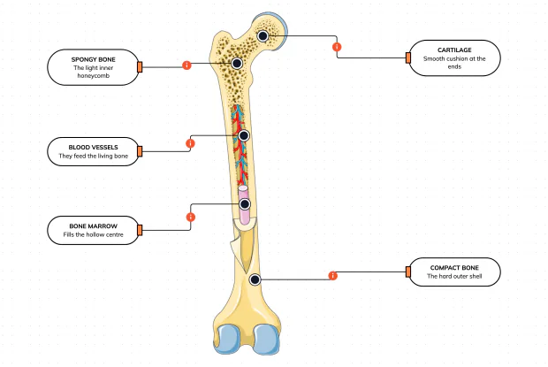
What Is a Bone Made Of?
Cut a bone open and it is nowhere near solid: a hard outer shell, a light honeycomb inside, a hollow centre full of marrow, blood vessels threading through, and a smooth cushion at each end. Learn what each part does — then label them yourself in an interactive playground.
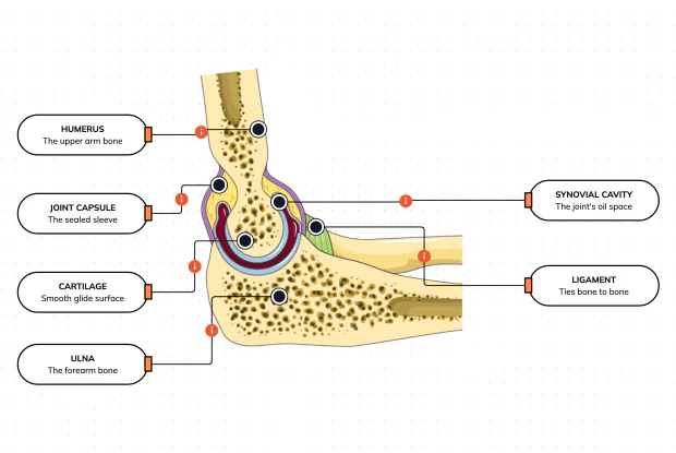
What Is Inside Your Elbow Joint?
A joint is where two bones meet and move — and it is far more than just the bones. Cut open the elbow and you find smooth cartilage caps, a sealed capsule of slippery fluid, and tough ligaments holding it all together. Learn what each part does, then label them yourself in an interactive playground.
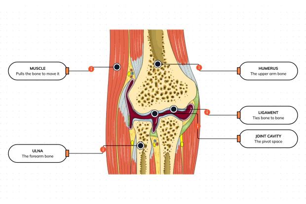
What Makes Your Elbow Bend?
Bones and joints are just the hardware; nothing moves until a muscle pulls. Label the elbow to see the parts that turn a joint into movement — the muscle that pulls, the ligament that holds bone to bone, the two bones it links, and the space they pivot in — then build it yourself in an interactive playground.
What Holds Your Elbow Together?
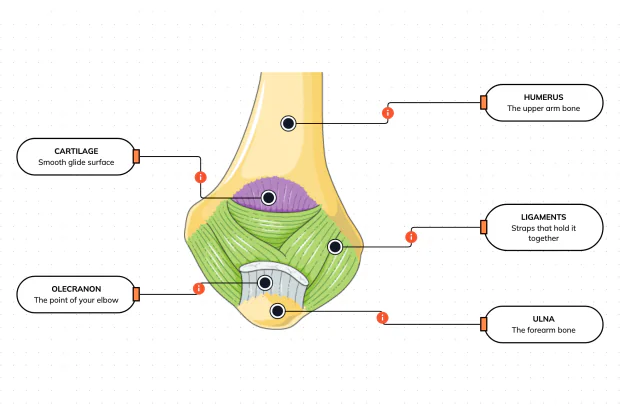
Your elbow is pulled on all day — by gravity, by every bag you carry, by every time you hang or push. So what stops the two bones from simply coming apart? Look at the back of the elbow and label the ligaments, the bony point, the cartilage and the two bones — and learn what holds the whole joint together.
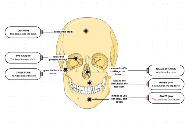
What Are the Main Bones of the Skull?
The skull looks like a single bone, but it is really about twenty-two locked together — with just one left free to move. Learn what each main part does, then label a skull yourself in an interactive playground.
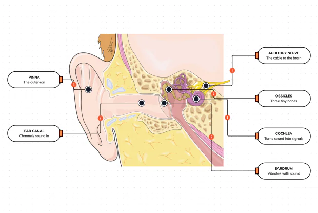
What Are the Parts of Your Ear?
Hearing is a chain of parts passing a sound along, each handing it to the next. Label the pinna, ear canal, eardrum, ossicles, cochlea and auditory nerve, and follow a sound all the way from the air outside to a signal your brain can read — in an interactive playground.
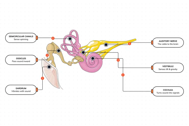
How Does Your Ear Keep You Balanced?
Your ear does a second, hidden job: balance. Inside it sit fluid-filled loops that sense spinning and a chamber that senses tilt and gravity. Label the semicircular canals, vestibule, cochlea, ossicles, eardrum and nerve, and learn how one organ both hears and keeps you upright — in an interactive playground.
What Are the Parts of the Eye You Can See?
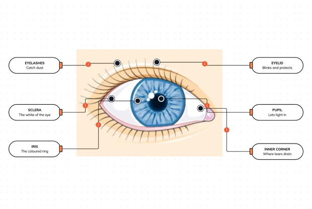
Before you look inside the eye, learn the parts you can already see in a mirror. Label the pupil, iris, sclera, eyelid, eyelashes and the tear-duct corner, and learn what each visible part does to let in light and protect the eye — in an interactive playground.
Where Do Your Tears Come From and Go?
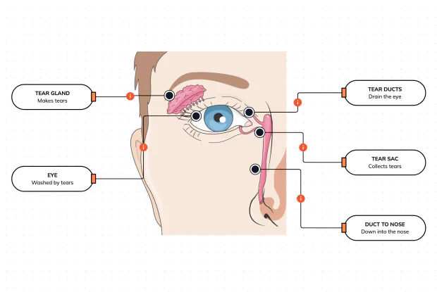
Your eyes make and drain tears all day to stay clean and wet. Label the tear gland, the eye, the tear ducts, the tear sac and the duct to the nose, and follow a tear from where it is made to where it drains — in an interactive playground.
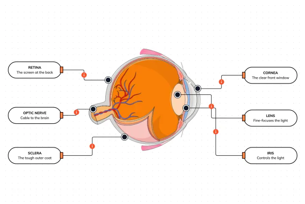
How Does Your Eye Turn Light Into a Picture?
Your eye works like a living camera. Cut it open and label the cornea, lens, iris, retina, optic nerve and sclera, and follow light as it is focused onto a screen at the back of the eye and sent to the brain — in an interactive playground.