How Does Your Eye Turn Light Into a Picture?
Root Concept
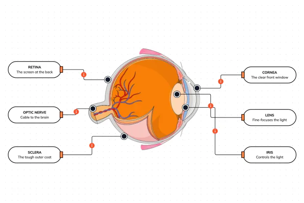
The eye works like a camera. Light enters through the clear cornea, passes the pupil (sized by the iris), and is focused by the lens onto the retina — the light-sensitive screen lining the back of the eye. The retina turns the image into signals that the optic nerve carries to the brain, and the tough sclera wraps and protects the whole eyeball.
CodePLU Goal
Upgrading Human Mental Models
Learn how to think in Workflows

Concept Development By codeplu.com
The inside of the eye, each part joined to the job it does as light is focused into a picture and sent to the brain
How Does Your Eye Turn Light Into a Picture?
Your eye is, quite literally, a living camera. It has a window at the front to let light in, lenses to focus that light, an adjustable opening to control the brightness, and a light-sensitive screen at the back to catch the picture — the same parts, doing the same jobs, as the camera in your phone. Once you see it that way, the inside of the eye stops being a jumble of odd names and becomes a simple chain: catch the light, focus it, land it on the screen, and send the picture to the brain.
Light begins its journey at the front. First it passes through the cornea, the clear domed window at the very front of the eye, which is not just a cover — its curved shape bends the incoming light and does most of the focusing. Just behind it, the iris (the coloured ring you can see from outside) opens and closes the pupil to let in the right amount of light. Then the light passes through the lens, a clear, flexible disc that fine-tunes the focus, changing its shape to sharpen things whether they are near or far.
The focused light then crosses the jelly-filled inside of the eyeball and lands on the retina — a delicate, light-sensitive screen lining the whole back wall, covered in tiny cells that turn the light into electrical signals. Those signals leave the eye through the optic nerve, the thick cable at the back that carries the picture to the brain, where you actually 'see' it. Wrapping and protecting the entire ball is the tough white sclera. In the playground above you will label all six parts and trace light's path from window to brain.
What Does Each Part of the Eye Do?
What does the cornea do?
The cornea is the clear, curved dome right at the front of the eye — the very first thing light passes through. People often think focusing is all done by the lens, but in fact the cornea does most of it. Because it is strongly curved and sits where light first enters, its shape bends the incoming rays and starts steering them toward a point at the back of the eye. Unlike the lens, the cornea's focusing power is fixed — it cannot change shape — so it does the bulk, steady focusing and leaves the fine adjustments to the lens. It has to be perfectly clear to let light through, which is why it has no blood vessels (they would block the view) and is kept moist and clean by your tears and blinking.
What does the lens do — and how is it different from the cornea?
The lens is the clear, flexible disc sitting just behind the iris and pupil, and its job is to fine-focus the light that the cornea has already started bending, landing it sharply on the retina. The key difference is that the lens can change shape: tiny muscles around it squeeze it fatter or let it go thinner, and that changes its focusing power. This is how you can look up from a book to a distant hill and have both snap into focus — the lens is adjusting, a trick called accommodation. The cornea does the big, fixed focusing; the lens does the small, adjustable focusing. As people get older the lens slowly stiffens and cannot change shape as easily, which is why so many people need reading glasses in middle age.
What is the iris doing here?
The iris is the coloured ring you already met from the outside of the eye — and here, in cross-section, you can see it sitting just in front of the lens. Its job is to control how much light gets in by changing the size of the pupil, the opening in its centre. In bright light it closes the pupil down to a small hole so the eye is not dazzled; in dim light it opens the pupil wide to gather more light. It works exactly like the aperture of a camera. Placing it right in front of the lens is no accident: light must pass through the pupil before it reaches the lens, so the iris sets the brightness first, and then the lens focuses whatever light it has let through.
What is the retina?
The retina is the eye's screen — a thin, delicate layer lining the whole inside of the back of the eyeball, and it is where the picture is actually formed. It is packed with millions of tiny light-sensitive cells that react when light lands on them, turning the focused image into a pattern of electrical signals. In a camera this is the job of the sensor (or, long ago, the film): the lens throws a focused image onto it, and it records what arrives. One curious fact is that the image lands on the retina upside-down, because of the way lenses bend light — and your brain simply flips it the right way up so the world looks normal. The retina is also where the blood vessels you can see branching across the back of the eye feed the hungry light-sensing cells.
What does the optic nerve do?
The optic nerve is the thick cable that leaves the back of the eyeball and runs to the brain, and it carries the signals the retina has made. On its own the retina cannot 'see' anything — it only creates the signals; seeing happens in the brain, and the optic nerve is the wire that delivers the message. Every one of the retina's millions of signals is bundled into this single nerve and sent to the vision centre at the back of the brain, which assembles them into the picture you experience. The one spot where the optic nerve joins the retina has no light-sensing cells, so it cannot detect light at all — that is your 'blind spot', a real gap in each eye's view that your brain quietly fills in so you never notice it.
What is the sclera?
The sclera is the tough, white outer coat that wraps almost the entire eyeball — the same white you can see from the front, shown here as the strong outer wall running all the way around. Its job is protection and shape: the eye is a soft, fluid-filled ball, and the sclera is the firm case that guards the delicate inner parts and holds the whole thing in its round form so the optics stay lined up. It also gives the muscles that move the eye something solid to attach to and pull on. At the very front it becomes clear and bulges out as the cornea — so in a sense the cornea is just the sclera's transparent window, letting light into the strong case that protects everything behind it.
Real World Example
Why Do So Many People Need Glasses?
Seeing clearly depends on the light being focused to land precisely on the retina. When it lands slightly in front of or behind the retina, the world looks blurry — and that small error is what glasses correct:
When the picture lands in the wrong place
The cornea, lens and retina
For you to see something sharply, the cornea and lens must bend the light so that it comes to a perfect focus exactly on the retina at the back of the eye. If everything matches up, the image is crisp. But the focus point depends on two things: how strongly the cornea and lens bend the light, and how long the eyeball is from front to back. If those two are even slightly mismatched, the light focuses a little in front of the retina or a little behind it, and by the time it actually reaches the screen it has blurred out again. The picture is being formed — just not in the right spot. This tiny mismatch, not any 'weakness' of the eye, is what most blurry vision really is.
Short sight and long sight
The shape of the eyeball
The two most common problems are opposite versions of that mismatch. In short-sightedness, the eyeball is a little too long (or the cornea too curved), so light focuses just in front of the retina — distant things blur while close things stay clear, which is why short-sighted people can read a book but not a road sign. In long-sightedness, the eyeball is a little too short, so light has not yet come to focus when it reaches the retina — close things blur while distant things are clearer. In both cases every part of the eye is healthy and working; the picture is simply landing a few millimetres off the screen. It is a plumbing problem of distances, not a broken part.
How glasses fix it
An added lens
Because the problem is just where the light comes to focus, the fix is beautifully simple: put another lens in front of the eye to nudge the focus point back onto the retina. Glasses and contact lenses do exactly this. A lens for short sight spreads the light out a little before it enters the eye, so the eye's own focus lands a bit further back — right on the retina. A lens for long sight does the opposite, bending the light inward a little so it focuses sooner. The eye's parts are left completely unchanged; the extra lens simply pre-adjusts the light so the cornea and lens land it in the right place. It is the same reason a camera has adjustable focus — and why understanding the eye's optics explains something almost everyone experiences.
FAQs
Final Words
The inside of the eye makes sense the moment you treat it as a camera. Light enters through the clear cornea, which does most of the focusing; the iris sets the brightness by sizing the pupil; the lens fine-focuses the image; and it lands on the retina, the light-sensitive screen at the back. The retina turns the picture into signals, the optic nerve carries them to the brain, and the tough sclera wraps and protects the whole ball. Catch the light, focus it, land it on the screen, send it to the brain.
Seeing this chain also explains everyday life — why almost everyone needs glasses (the focus landing a fraction off the retina), why you have a blind spot, and why the world does not look upside-down even though the image is. You have now met the eye from the outside and the inside; next you will see the muscles around it that aim this remarkable camera wherever you want to look.
Continue This Track
This concept is part 10 of The Human Body, Head to Toe.
What Is a Bone Made Of?
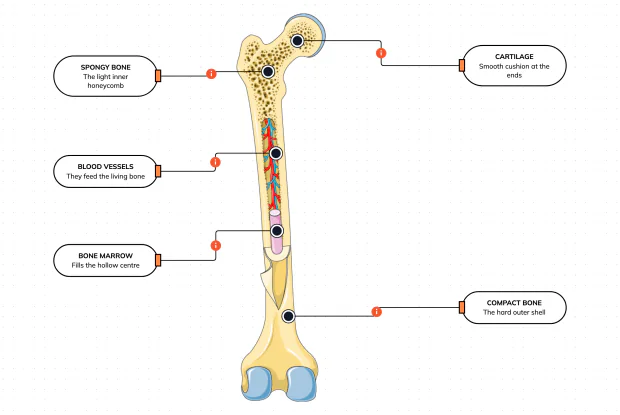
Cut a bone open and it is nowhere near solid: a hard outer shell, a light honeycomb inside, a hollow centre full of marrow, blood vessels threading through, and a smooth cushion at each end. Learn what each part does — then label them yourself in an interactive playground.
What Is Inside Your Elbow Joint?
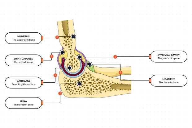
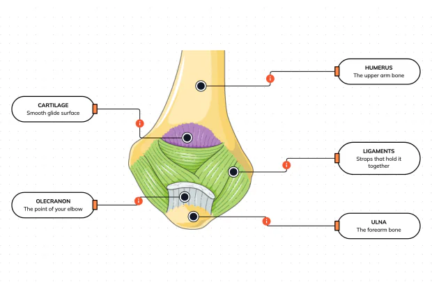
A joint is where two bones meet and move — and it is far more than just the bones. Cut open the elbow and you find smooth cartilage caps, a sealed capsule of slippery fluid, and tough ligaments holding it all together. Learn what each part does, then label them yourself in an interactive playground.
What Makes Your Elbow Bend?
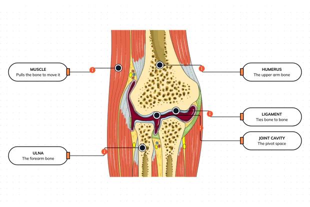
Bones and joints are just the hardware; nothing moves until a muscle pulls. Label the elbow to see the parts that turn a joint into movement — the muscle that pulls, the ligament that holds bone to bone, the two bones it links, and the space they pivot in — then build it yourself in an interactive playground.
What Holds Your Elbow Together?
Your elbow is pulled on all day — by gravity, by every bag you carry, by every time you hang or push. So what stops the two bones from simply coming apart? Look at the back of the elbow and label the ligaments, the bony point, the cartilage and the two bones — and learn what holds the whole joint together.
What Are the Main Bones of the Skull?
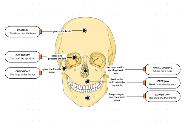
The skull looks like a single bone, but it is really about twenty-two locked together — with just one left free to move. Learn what each main part does, then label a skull yourself in an interactive playground.
What Are the Parts of Your Ear?
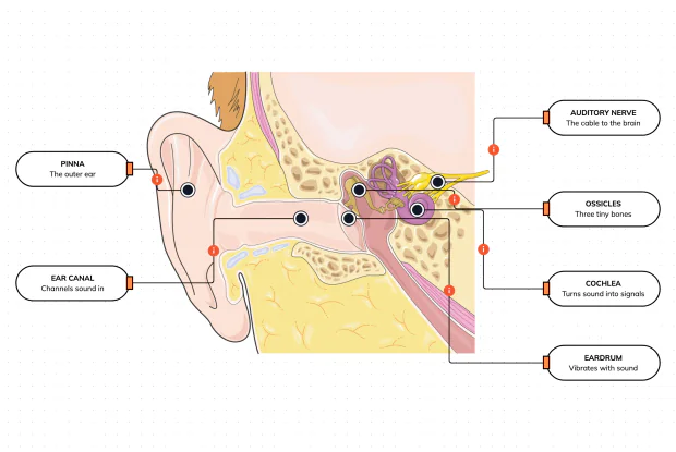
Hearing is a chain of parts passing a sound along, each handing it to the next. Label the pinna, ear canal, eardrum, ossicles, cochlea and auditory nerve, and follow a sound all the way from the air outside to a signal your brain can read — in an interactive playground.
How Does Your Ear Keep You Balanced?
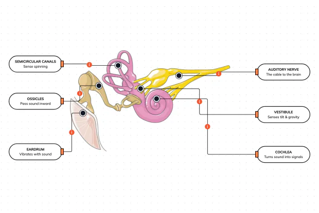
Your ear does a second, hidden job: balance. Inside it sit fluid-filled loops that sense spinning and a chamber that senses tilt and gravity. Label the semicircular canals, vestibule, cochlea, ossicles, eardrum and nerve, and learn how one organ both hears and keeps you upright — in an interactive playground.
What Are the Parts of the Eye You Can See?
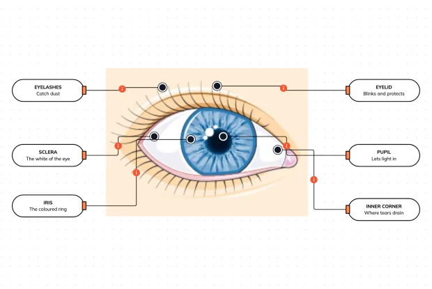
Before you look inside the eye, learn the parts you can already see in a mirror. Label the pupil, iris, sclera, eyelid, eyelashes and the tear-duct corner, and learn what each visible part does to let in light and protect the eye — in an interactive playground.
Where Do Your Tears Come From and Go?
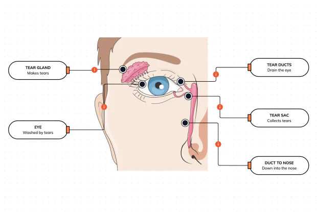
Your eyes make and drain tears all day to stay clean and wet. Label the tear gland, the eye, the tear ducts, the tear sac and the duct to the nose, and follow a tear from where it is made to where it drains — in an interactive playground.
How Does Your Eye Turn Light Into a Picture?
Your eye works like a living camera. Cut it open and label the cornea, lens, iris, retina, optic nerve and sclera, and follow light as it is focused onto a screen at the back of the eye and sent to the brain — in an interactive playground.