What Does the Back of Your Eye Look Like?
Root Concept
The retina is the light-sensitive screen lining the back of the eye. It is not uniform: the fovea, at the centre of the macula, gives the sharpest vision; the optic disc, where the nerve and blood vessels enter, has no light sensors and is the blind spot; and blood vessels spread across the surface to feed the light-sensing cells.
CodePLU Goal
Upgrading Human Mental Models
Learn how to think in Workflows

Concept Development By codeplu.com
The back of the eye, each part joined to the job it does — where the retina sees sharpest, where it is blind, and how it is fed
Why Can You Only See Sharply Straight Ahead?
Here is something strange you can test right now: keep your eyes fixed on this word, and without moving them, try to read the words a few lines above or below. You can't — they are blurry, even though your eye is wide open and pointing forward. Only the very thing you look straight at is sharp; everything around it is surprisingly rough. That is not a fault in your eyes. It is because the retina, the screen at the back of your eye, is not the same all over — and this view of the back of the eye shows exactly why.
Almost the whole back of the eye is covered by the retina, the light-sensitive screen packed with cells that turn light into signals. But look closely and you can see two special spots. On one side is a darker patch called the macula, and at its very centre a bright point called the fovea — this tiny area is crammed with far more light-sensing cells than anywhere else, and it is where your vision is razor-sharp. Everywhere else on the retina has fewer sensors, so it gives you only rough, side-of-the-eye vision, good for spotting movement but not detail.
On the other side is a bright pale circle, the optic disc — the spot where the optic nerve leaves the eye and the blood vessels enter and branch out across the retina. It is the eye's cable-and-plumbing hub, but it has one odd consequence: because that spot is taken up by the nerve and vessels, it has no light sensors at all, making it a real blind spot in your vision. In the playground above you will label the retina, the macula and fovea, the optic disc, and the vessels — and the label on each connection tells you the job that part does.
What Is Each Part of the Retina For?
What is the retina?
The retina is the light-sensitive layer that lines the inside of the back of the eye — the screen the lens throws its image onto. It is covered in millions of tiny cells that react to light and turn the focused image into electrical signals, which then travel to the brain. If the eye is a camera, the retina is its sensor. What this view shows, that a side-on diagram cannot, is that the retina is a real surface with features on it — it is not a smooth, uniform screen but has special regions that do different things, plus the vessels that keep it alive. Everything else labelled here is a particular part of this one light-catching sheet.
What is the optic disc — and why is it your blind spot?
The optic disc is the bright, pale circle you can see to one side of the retina, and it is the eye's hub: the place where the optic nerve gathers up all the retina's signals and leaves the eye toward the brain, and where the main blood vessels enter and fan out. But there is a catch. Because that whole patch is filled with nerve fibres and vessels leaving and entering, there is no room for light-sensing cells there — so that spot cannot detect light at all. This is your blind spot: a genuine hole in the vision of each eye. You never notice it because your two eyes cover for each other, and because your brain quietly fills in the gap with a guess of what should be there.
What is the macula?
The macula is the small, slightly darker patch near the centre of the retina — the region directly in line with whatever you are looking straight at. It is packed with far more light-sensing cells than the outer retina, and in particular the kind that see fine detail and colour, so the macula is responsible for your sharp, detailed, colourful central vision: reading, recognising faces, threading a needle. The rest of the retina handles your side (peripheral) vision, which is much rougher and nearly colourless but good at detecting movement. This is why you turn to look directly at something you want to see clearly — you are swinging its image onto the macula. When the macula is damaged, as in macular degeneration, people lose their sharp central sight while keeping their side vision.
What is the fovea, and why does it matter so much?
The fovea is the tiny bright pit at the very centre of the macula, and it is the single most important point on the whole retina: the spot where your vision is sharpest of all. It is packed most densely with the detail-and-colour cells and has almost nothing else in the way, so the light lands on the sensors directly. When you look straight at something, you are aiming its image onto this pinpoint — you are reading these words with your fovea right now. It is astonishingly small, which is exactly why only the thing you look directly at is truly sharp: only a tiny patch of the world falls on the fovea at any moment. That is also why your eyes constantly flick around — to bring one detail after another onto this one sharp spot.
Why are there blood vessels all over the retina?
The branching red vessels spreading across the retina are there because the retina's light-sensing cells are extremely hungry — detecting light and firing signals non-stop takes a lot of energy, and that means a constant supply of blood carrying oxygen and nutrients. The vessels enter at the optic disc and spread out to reach the whole surface. Interestingly, they avoid crossing the fovea itself, so that nothing blocks the light where vision is sharpest. The retina is the one place in the entire body where a doctor can look at your blood vessels directly, without any cutting — which is why an eye examination can reveal signs of conditions like diabetes and high blood pressure, which damage these tiny vessels early.
Real World Example
Why Do You Have to Move Your Eyes to Read a Single Line?
Because the retina sees sharply in only a tiny area and is blind in another, your everyday vision works in ways that make perfect sense once you know the back of the eye:
Why you move your eyes to read
The fovea
Only the fovea sees fine detail, and it is tiny — it takes in just a few letters at a time at reading distance. That is why you cannot simply hold your eyes still and absorb a whole page; you have to move them, flicking the fovea from word to word so each one, in turn, lands on that single sharp spot. Everything on the page not currently on your fovea is genuinely blurry, as the experiment of fixing your gaze on one word proves. It feels as though your whole field of view is sharp, but that is an illusion: your eyes dart around so fast, and your brain stitches the sharp snapshots together so smoothly, that you never notice you are really seeing the world clearly through a pinhole.
Finding your blind spot
The optic disc
You can actually find the blind spot the optic disc creates. Close your right eye, hold up two fingers apart at arm's length, stare at the right finger with your left eye, and slowly move the left finger outward — at a certain point it vanishes, then reappears as you keep moving. For that moment its image is falling on the optic disc, where there are no light sensors, so it simply disappears. What is remarkable is that you do not see a black hole where it went — your brain fills the gap with the surrounding orange, or pattern, or colour, so convincingly that you never notice the blind spot in normal life. It is a vivid demonstration that what you 'see' is partly the brain's best guess.
Why a doctor shines a light in your eye
The retina and its vessels
When a doctor shines a bright light into your eye and peers in, they are looking at this exact view — the retina and its blood vessels. The back of the eye is the only place in the whole body where blood vessels can be seen directly, in the living person, without any surgery. That makes the retina a unique window on your general health: diseases that damage small blood vessels, such as diabetes and high blood pressure, often show their earliest signs here, in changes to the retinal vessels or the retina itself. So an eye exam is not only about how well you see — it can catch conditions affecting the whole body, spotted through the one clear porthole the eye provides.
FAQs
Final Words
The retina is the screen at the back of your eye, but the great surprise is that it is not the same all over. At the fovea, in the centre of the macula, it is crammed with sensors and your vision is razor-sharp; across the rest it is rougher, giving only side vision; and at the optic disc, where the nerve and vessels pass through, it is completely blind. Blood vessels spread over the whole surface to feed its hungry cells. One screen, but with a sharp spot, a rough surround, and a blind hole.
This uneven design explains everyday life — why you move your eyes to read, why you have a blind spot you never notice, and why a doctor can read your general health from the vessels here. You have now followed sight from the outside of the eye, through its focusing parts, right onto the retina where seeing begins. Next you can look at how the eye keeps that image sharp — what 'normal vision' really means.
Continue This Track
This concept is part 11 of The Human Body, Head to Toe.
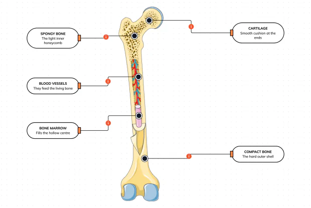
What Is a Bone Made Of?
Cut a bone open and it is nowhere near solid: a hard outer shell, a light honeycomb inside, a hollow centre full of marrow, blood vessels threading through, and a smooth cushion at each end. Learn what each part does — then label them yourself in an interactive playground.
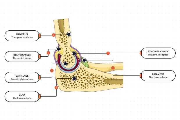
What Is Inside Your Elbow Joint?
A joint is where two bones meet and move — and it is far more than just the bones. Cut open the elbow and you find smooth cartilage caps, a sealed capsule of slippery fluid, and tough ligaments holding it all together. Learn what each part does, then label them yourself in an interactive playground.
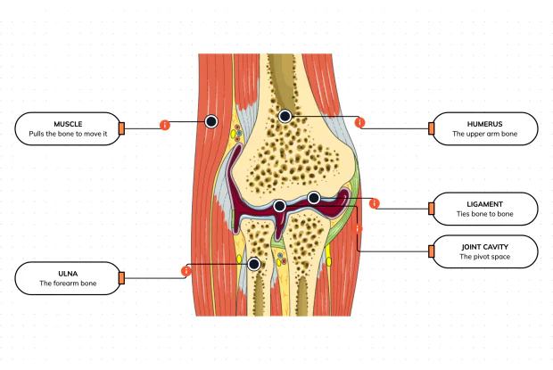
What Makes Your Elbow Bend?
Bones and joints are just the hardware; nothing moves until a muscle pulls. Label the elbow to see the parts that turn a joint into movement — the muscle that pulls, the ligament that holds bone to bone, the two bones it links, and the space they pivot in — then build it yourself in an interactive playground.
What Holds Your Elbow Together?
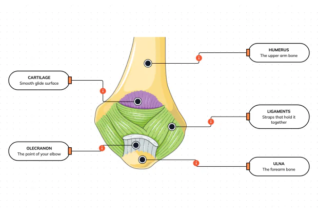
Your elbow is pulled on all day — by gravity, by every bag you carry, by every time you hang or push. So what stops the two bones from simply coming apart? Look at the back of the elbow and label the ligaments, the bony point, the cartilage and the two bones — and learn what holds the whole joint together.
What Are the Main Bones of the Skull?
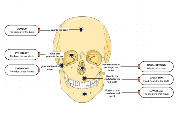
The skull looks like a single bone, but it is really about twenty-two locked together — with just one left free to move. Learn what each main part does, then label a skull yourself in an interactive playground.
What Are the Parts of Your Ear?
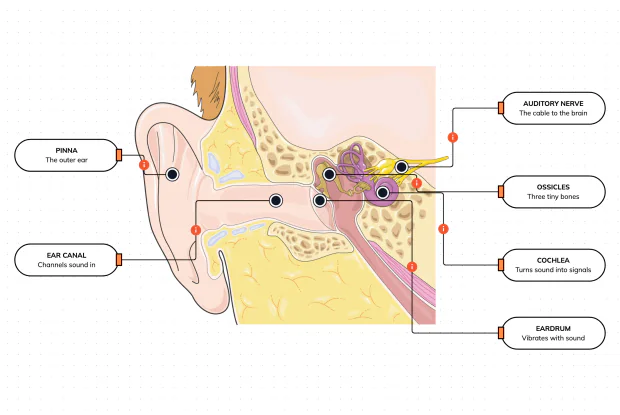
Hearing is a chain of parts passing a sound along, each handing it to the next. Label the pinna, ear canal, eardrum, ossicles, cochlea and auditory nerve, and follow a sound all the way from the air outside to a signal your brain can read — in an interactive playground.
How Does Your Ear Keep You Balanced?
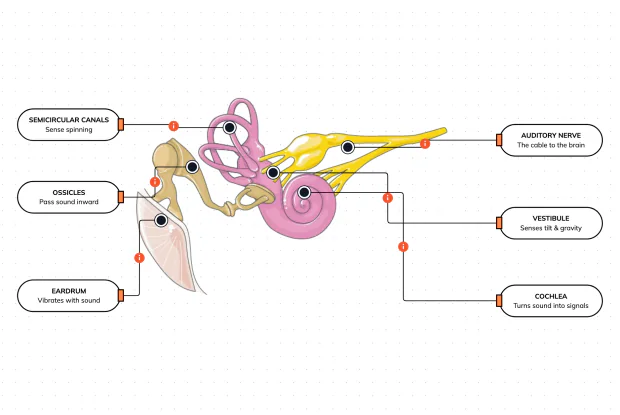
Your ear does a second, hidden job: balance. Inside it sit fluid-filled loops that sense spinning and a chamber that senses tilt and gravity. Label the semicircular canals, vestibule, cochlea, ossicles, eardrum and nerve, and learn how one organ both hears and keeps you upright — in an interactive playground.
What Are the Parts of the Eye You Can See?
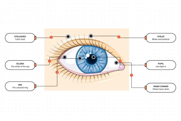
Before you look inside the eye, learn the parts you can already see in a mirror. Label the pupil, iris, sclera, eyelid, eyelashes and the tear-duct corner, and learn what each visible part does to let in light and protect the eye — in an interactive playground.
Where Do Your Tears Come From and Go?
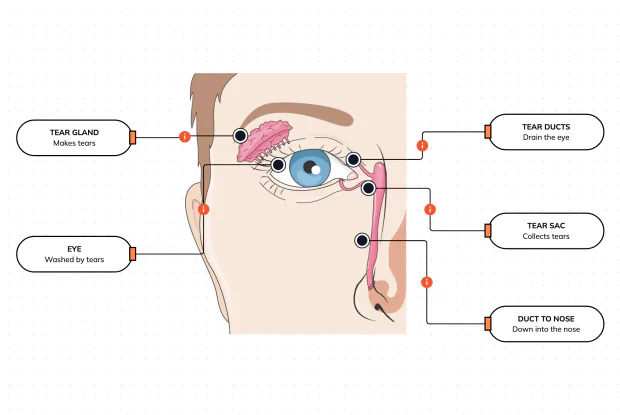
Your eyes make and drain tears all day to stay clean and wet. Label the tear gland, the eye, the tear ducts, the tear sac and the duct to the nose, and follow a tear from where it is made to where it drains — in an interactive playground.
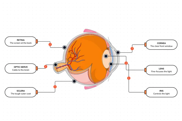
How Does Your Eye Turn Light Into a Picture?
Your eye works like a living camera. Cut it open and label the cornea, lens, iris, retina, optic nerve and sclera, and follow light as it is focused onto a screen at the back of the eye and sent to the brain — in an interactive playground.