How Does Your Body Hold and Release Waste?
Root Concept
The rectum is the storage chamber at the end of the gut. Its stretchy folded wall expands as waste collects and signals the urge to go. Waste leaves through the short anal canal, guarded by a ring of sphincter muscle — an inner, automatic sphincter and an outer, voluntary one — while the anal columns are cushioned ridges of lining at the top of the canal.
CodePLU Goal
Upgrading Human Mental Models
Learn how to think in Workflows

Concept Development By codeplu.com
The rectum and anal canal, each part joined to the job it does — storing waste and controlling exactly when it is released
How Do You Decide When to Go to the Toilet?
The very end of the digestive tract solves a surprisingly tricky problem: waste is ready to leave at times that are rarely convenient, so the body needs a way to store it and release it only when you choose. That is the job of the rectum and the anal canal. It is a storage chamber with a controllable exit — and the control is the clever part, because it lets you feel the need to go, then hold on until you reach a toilet. Almost every one of us relies on this system countless times without a thought, and learning to control it is one of the first things we master as toddlers.
The rectum itself is the storage chamber — the stretchy maroon cavity that makes up most of this picture. Waste arriving from the sigmoid colon collects here, and the folded rectal wall expands to hold it. As it stretches, it sends signals to the brain: this is the feeling of needing to go. If now is not a good time, the walls relax to accommodate a bit more, and the urge eases off for a while.
At the bottom, the rectum narrows into the short anal canal, the final passage to the outside, lined at its top with cushioned ridges called the anal columns. Wrapped around the canal is the crucial part: rings of sphincter muscle that hold the exit tightly shut and relax only when you allow it. In the playground above you will label the rectum, rectal wall, anal columns, anal canal and sphincter muscle — and the label on each connection tells you the job that part does.
What Are the Parts of the Rectum and Anal Canal?
What does the rectum do?
The rectum is the storage chamber at the end of the large intestine — the wide maroon cavity that dominates this figure. Its job is to hold solid waste (faeces) until it is convenient to pass it. For most of the time the rectum is actually empty; waste is stored higher up, in the colon, and only moves down into the rectum when it is nearly time to go. When it does arrive, the rectum holds it and, as we will see, raises the alarm. This storage role is what makes controlled, occasional trips to the toilet possible, rather than a constant dribble — the rectum lets waste accumulate and be released in one go, at a chosen moment. It is the buffer between the steady work of the gut and your convenience.
How does the rectal wall know when it's full?
The rectal wall is folded and stretchy, and that stretchiness is the key to the whole system. When waste moves down into the rectum, the wall is stretched, and special stretch-sensors in it detect that expansion and fire off signals to the spinal cord and brain. Your brain reads those signals as the familiar feeling of needing to go — the fuller the rectum, the stronger the urge. Cleverly, if you decide it is not the moment, the wall can relax and expand a little more to make room, and the urge temporarily fades, buying you time. So the rectal wall is not just a passive bag; it is a sensor that measures how full it is and tells your brain, which is how you become aware of the need at all. Without this signal, you would never know it was time.
What controls when waste is released?
The exit is guarded by the sphincter muscle — and there are actually two rings, working as a two-lock system. The inner ring (the internal sphincter) is made of the gut's own smooth muscle and is automatic: it stays contracted on its own and relaxes by reflex when the rectum fills, without you deciding. The outer ring (the external sphincter) is made of muscle you control voluntarily, like the muscles in your arm — you can squeeze it shut or let it relax at will. This pairing is what gives you control: when the rectum fills and the inner, automatic lock relaxes, the outer, voluntary lock is still shut because you are holding it, so nothing escapes until you choose to relax it too. Two locks, one automatic and one under your command, are the reason you can feel the urge yet wait for the right time and place.
What is the anal canal?
The anal canal is the short final passage — only a few centimetres long — leading from the rectum to the outside of the body. It is the last section of the entire digestive tract, the tube that began all the way back at your mouth. Its walls are the sphincter muscles that keep it closed, so most of the time the canal is squeezed shut, sealing the exit; it opens only briefly to let waste pass. Because it is the boundary between inside and outside, the lining changes along the canal from the soft inner gut lining to tougher, more skin-like tissue near the opening, which can sense touch and help tell the difference between solid, liquid and gas — a subtle but useful ability. The anal canal is short and simple, but it is the guarded doorway at the very end of the gut.
What are the anal columns?
At the top of the anal canal, the lining is raised into a ring of pale, finger-like ridges called the anal columns, which you can see as the pale vertical folds in the figure. These columns are cushions: each contains a small network of veins, and together they act like soft, blood-filled pads that help the anal canal seal completely shut. A muscle ring alone might not make a perfect seal, but these squashy cushions fill the last small gaps, helping to keep in gas and liquid as well as solids. They are useful — but they are also the structures that cause a very common problem: if the veins inside them swell and enlarge (from straining, pregnancy or sitting too long), the cushions become hemorrhoids (piles), which can itch, hurt or bleed. So the anal columns are the sealing cushions of the exit, and their vein-filled nature is exactly why hemorrhoids happen where they do.
Real World Example
Why Can You 'Hold It' Until You Reach a Toilet?
The rectum and anal canal are built around one goal: letting you control exactly when you go. Three features make that possible — and explain some very common experiences:
Two locks on one door
The internal and external sphincters
The exit has two rings of muscle, and the difference between them is everything. The inner ring, the internal sphincter, is automatic: it stays shut by itself and relaxes by reflex when the rectum fills — you have no direct control over it. The outer ring, the external sphincter, is voluntary: it is under your conscious command, like clenching a fist. So when the rectum fills and the automatic lock reflexively opens, the door does not swing open, because the voluntary lock is still held shut by you. This two-lock design is precisely what lets you feel the urge yet decide the moment: the automatic sphincter says 'ready', but the voluntary one waits for your say-so. Learning to control that voluntary muscle is exactly what toilet training a toddler is, and losing that control is what causes incontinence.
The stretch that tells you 'go'
The rectal wall
You can only decide to hold on if you know there is something to hold — and that awareness comes from the stretchy rectal wall. As waste moves into the rectum and stretches the wall, sensors in it signal your brain, producing the urge to go, growing stronger as the rectum fills. If you choose to wait, the wall relaxes to make more room and the urge subsides for a while, which is why the feeling comes in waves rather than staying constant. This is also why ignoring the urge repeatedly can backfire: the rectum stretches and adapts, the signal weakens, waste sits longer and more water is drawn out of it, making constipation more likely. The rectal wall's stretch signal is the alarm bell of the whole system — without it, you would have no warning at all.
Why the cushions can swell
The anal columns and hemorrhoids
The anal columns are cushions of vein-filled tissue that help seal the canal shut — a useful part of the design, but a vulnerable one. The small veins inside them can become swollen and enlarged under pressure: straining to pass hard stool, long periods of sitting, or the pressure of pregnancy can all engorge these veins until the cushions balloon into hemorrhoids (piles). Because the cushions sit right at the exit and are rich in blood vessels, swollen ones can itch, ache, or bleed a little — one of the most common minor complaints there is. It is a neat example of anatomy explaining a symptom: hemorrhoids happen exactly where they do because that is where the sealing cushions and their veins are, and they are made worse by anything that raises the pressure on those veins.
FAQs
Final Words
The rectum and anal canal are the controlled exit at the very end of your gut. The rectum stores waste, and its stretchy folded wall signals your brain when it is full — the urge to go. Waste leaves through the short anal canal, sealed by two rings of sphincter muscle: an inner, automatic one and an outer, voluntary one. The anal columns are cushioned, vein-filled ridges that help the canal seal completely. Storage, a fullness signal, and a two-lock exit — that is the whole system.
That two-lock design is the quiet reason civilised life is possible: you feel the need, but you decide the moment. It explains toilet training, incontinence, and even why hemorrhoids form where they do. With the rectum, the tour of the digestive tract is complete — you have followed food all the way from the mouth, through the stomach, the liver and pancreas, the long absorbing small intestine and the water-reclaiming large intestine, to this final, controllable door.
Continue This Track
This concept is part 40 of The Human Body, Head to Toe.
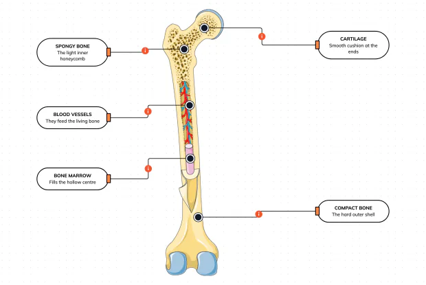
What Is a Bone Made Of?
Cut a bone open and it is nowhere near solid: a hard outer shell, a light honeycomb inside, a hollow centre full of marrow, blood vessels threading through, and a smooth cushion at each end. Learn what each part does — then label them yourself in an interactive playground.
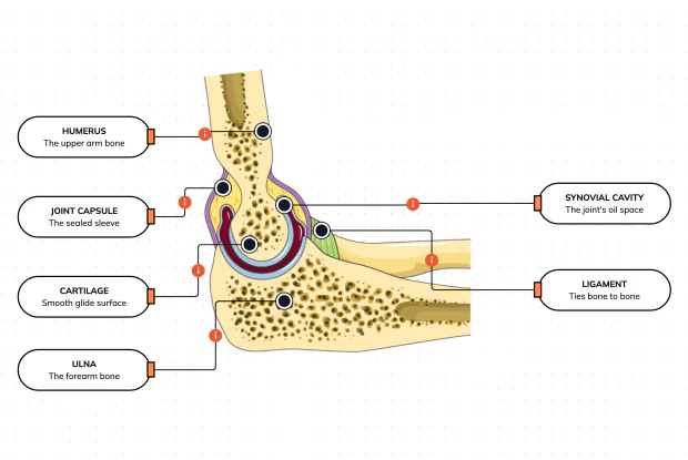
What Is Inside Your Elbow Joint?
A joint is where two bones meet and move — and it is far more than just the bones. Cut open the elbow and you find smooth cartilage caps, a sealed capsule of slippery fluid, and tough ligaments holding it all together. Learn what each part does, then label them yourself in an interactive playground.
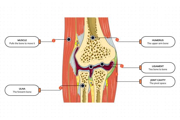
What Makes Your Elbow Bend?
Bones and joints are just the hardware; nothing moves until a muscle pulls. Label the elbow to see the parts that turn a joint into movement — the muscle that pulls, the ligament that holds bone to bone, the two bones it links, and the space they pivot in — then build it yourself in an interactive playground.
What Holds Your Elbow Together?
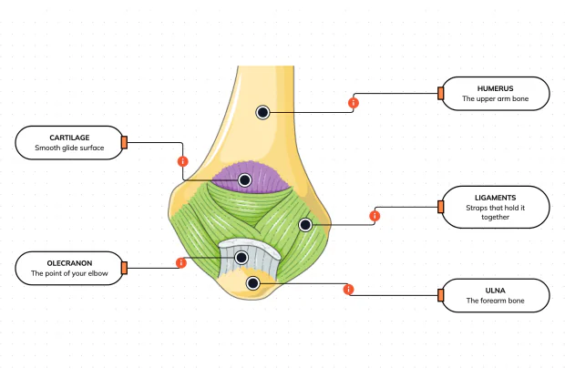
Your elbow is pulled on all day — by gravity, by every bag you carry, by every time you hang or push. So what stops the two bones from simply coming apart? Look at the back of the elbow and label the ligaments, the bony point, the cartilage and the two bones — and learn what holds the whole joint together.
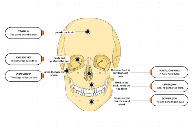
What Are the Main Bones of the Skull?
The skull looks like a single bone, but it is really about twenty-two locked together — with just one left free to move. Learn what each main part does, then label a skull yourself in an interactive playground.
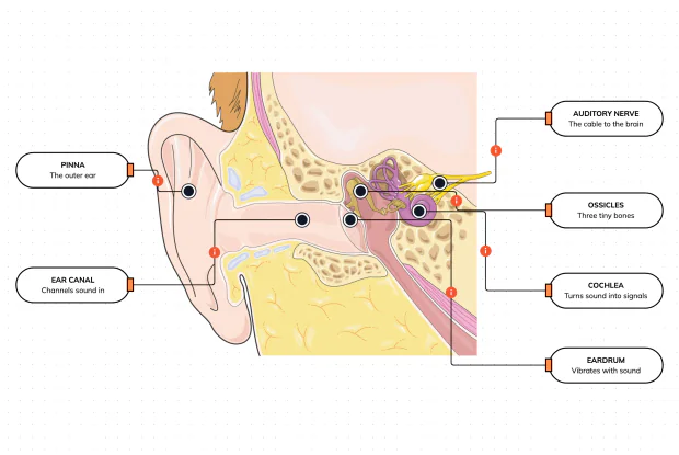
What Are the Parts of Your Ear?
Hearing is a chain of parts passing a sound along, each handing it to the next. Label the pinna, ear canal, eardrum, ossicles, cochlea and auditory nerve, and follow a sound all the way from the air outside to a signal your brain can read — in an interactive playground.
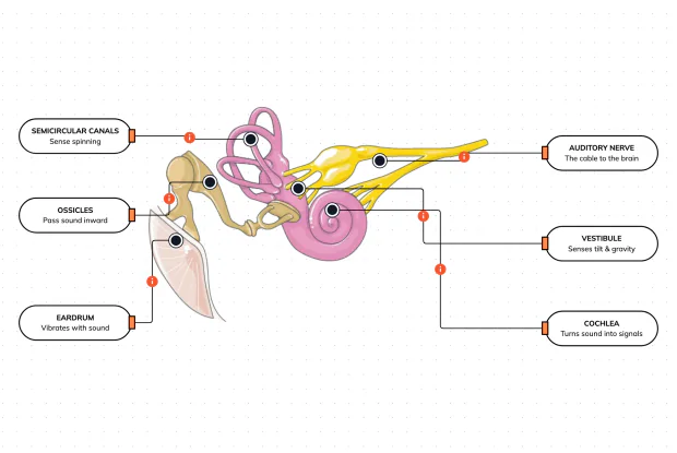
How Does Your Ear Keep You Balanced?
Your ear does a second, hidden job: balance. Inside it sit fluid-filled loops that sense spinning and a chamber that senses tilt and gravity. Label the semicircular canals, vestibule, cochlea, ossicles, eardrum and nerve, and learn how one organ both hears and keeps you upright — in an interactive playground.
What Are the Parts of the Eye You Can See?
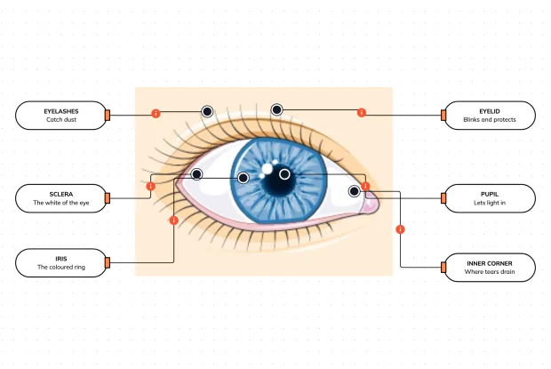
Before you look inside the eye, learn the parts you can already see in a mirror. Label the pupil, iris, sclera, eyelid, eyelashes and the tear-duct corner, and learn what each visible part does to let in light and protect the eye — in an interactive playground.
Where Do Your Tears Come From and Go?
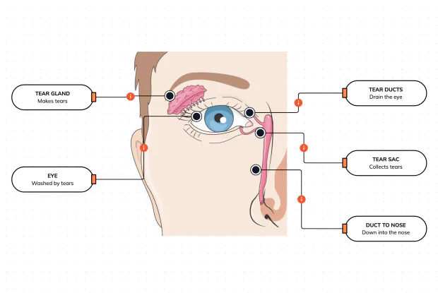
Your eyes make and drain tears all day to stay clean and wet. Label the tear gland, the eye, the tear ducts, the tear sac and the duct to the nose, and follow a tear from where it is made to where it drains — in an interactive playground.
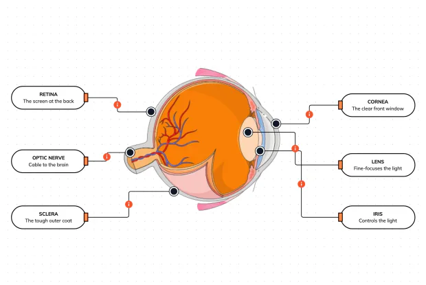
How Does Your Eye Turn Light Into a Picture?
Your eye works like a living camera. Cut it open and label the cornea, lens, iris, retina, optic nerve and sclera, and follow light as it is focused onto a screen at the back of the eye and sent to the brain — in an interactive playground.