What Does the Placenta Do?
Root Concept
The placenta is a temporary organ attached to the wall of the uterus that acts as a swap-point: the mother's blood and the baby's blood run close together inside it without ever mixing, so oxygen and food pass one way and waste passes back. The umbilical cord carries that supply to the baby, and the placenta normally sits high on the uterus wall, leaving the exit at the cervix clear.
CodePLU Goal
Upgrading Human Mental Models
Learn how to think in Workflows

Concept Development By codeplu.com
A baby's supply line — the placenta high on the uterus wall, the cord running down to the baby, and the cervix left clear below
How Does a Baby Eat and Breathe Before It Is Born?
A baby in the uterus does not eat, and its lungs are full of fluid rather than air. Everything it needs arrives ready-made through one connection: the umbilical cord, running from its navel up to a dark, spongy disc stuck to the wall of the uterus. That disc is the placenta, and it is the only organ a human body ever builds on purpose and then throws away.
The placenta grows from the same cells as the baby, not from the mother, and it plants itself into the uterus lining where the mother's blood supply is richest. Inside, the two blood supplies are brought within a hair's breadth of each other across a thin barrier. Oxygen and food cross from mother to baby; carbon dioxide and waste cross back the other way. The two bloods never actually mix, which is a detail worth holding on to.
Notice where the placenta sits in the picture: high up, across the top of the uterus, a long way from the narrow opening at the bottom. That is what 'normal location' means, and it matters — the cervix has to be free for the baby to come through at the end. In the playground you will label the placenta, the cord, the uterus wall, the fluid the baby floats in, and the cervix.
How Does the Placenta Actually Work?
Why do the two blood supplies never mix?
This is the part almost everyone gets wrong. It is easy to imagine the cord as a pipe plumbed straight into the mother, but the baby has its own entirely separate circulation. Inside the placenta, tiny branching tufts of the baby's blood vessels dangle into pools of the mother's blood, like fingers dipped in a bath. Only a very thin barrier separates the two. Oxygen, sugar, vitamins and antibodies slip across that barrier into the baby's vessels, while carbon dioxide and waste slip back into the mother's blood for her kidneys and lungs to clear. Keeping the bloods apart matters enormously: it lets the mother's immune system tolerate a baby whose blood type and genes are not her own.
What does the umbilical cord carry?
The cord is the baby's lifeline, and by the end of pregnancy it is about as long as the baby is tall and roughly as thick as a finger. Inside are three blood vessels, wound in a spiral and cushioned by a jelly that stops them kinking when the baby moves and tumbles. Two of them carry used, oxygen-poor blood from the baby back to the placenta to be cleaned and refreshed; one carries the refreshed, oxygen-rich blood back to the baby. Everything the baby takes in for nine months travels this one route — which is why the navel, the small scar where the cord was attached, is on every human body. It is the mark of where your supply line used to be.
Why does it matter where the placenta sits?
The placenta can attach almost anywhere on the inside of the uterus, and normally it lands high — on the top or upper wall, as in the picture — leaving the cervix at the bottom completely clear. That matters at the end of pregnancy, because the baby has to pass through the cervix to be born. If a placenta instead settles low down and covers the cervix, it blocks the exit, and because the placenta is packed with blood vessels it would be torn as labour opened the cervix. Doctors check the placenta's position on a routine scan for exactly this reason, and a low one usually means planning a caesarean birth. A high placenta is a quiet piece of good news.
Real World Example
Why the Placenta Is Delivered Too
Follow the placenta through the end of a pregnancy and three everyday things make sense:
The afterbirth
The organ leaves as well
Once a baby is born, the job is not quite finished. The uterus keeps contracting, the placenta peels away from the wall it was planted in, and a few minutes later it is delivered too — which is why it is often called the afterbirth. It comes out as a dark red disc, roughly the size of a dinner plate and about as heavy as a large book, with the cut cord trailing from it. Midwives examine it carefully to check that none has been left behind inside, because a fragment left in place can cause bleeding. It is the only time most people ever see an organ that their own body built and then finished with.
Cutting the cord
The supply line is closed
The moment a baby is born and takes its first breath, its lungs take over the job the placenta was doing, and the cord is no longer needed. The cord is clamped and cut, which the baby does not feel — there are no nerves in it. The short stump left on the baby dries out and drops off within a week or two, leaving the navel. Doctors now usually wait a minute or so before clamping so that blood still in the cord and placenta can drain into the baby, giving it a useful extra supply of iron. One organ hands over to another, and a temporary connection becomes a permanent scar.
What this tells you
A whole organ, built to be temporary
Nothing else in the body works this way. The placenta is grown from scratch for one pregnancy, takes over the work of the lungs, gut, kidneys and part of the hormone system for nine months, and is then discarded. That is why the position it chooses is checked on a scan, why its health is watched closely through pregnancy, and why problems with it affect the baby's growth so directly — if the swap-point is not working well, the baby's only supply route is restricted. Seeing it as a temporary organ rather than a piece of packaging is the shift that makes pregnancy biology click.
FAQs
Final Words
The placenta is a temporary organ built for one pregnancy. It plants itself in the wall of the uterus, brings the mother's blood and the baby's blood close together without letting them mix, and passes oxygen and food one way and waste the other. The umbilical cord carries that supply to the baby, the amniotic fluid cushions it, and the muscular uterus wall holds everything and does the pushing at the end.
Where the placenta lands matters as much as how it works: high on the wall leaves the cervix clear for birth, and that is exactly what a routine scan is checking. Next, look at how the baby itself lies in the last weeks before birth, and why being head-down makes the journey out so much easier.
Continue This Track
This concept is part 62 of The Human Body, Head to Toe.
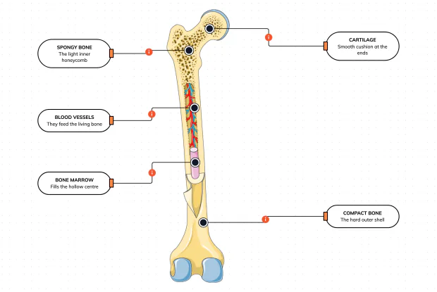
What Is a Bone Made Of?
Cut a bone open and it is nowhere near solid: a hard outer shell, a light honeycomb inside, a hollow centre full of marrow, blood vessels threading through, and a smooth cushion at each end. Learn what each part does — then label them yourself in an interactive playground.
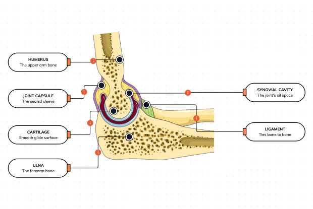
What Is Inside Your Elbow Joint?
A joint is where two bones meet and move — and it is far more than just the bones. Cut open the elbow and you find smooth cartilage caps, a sealed capsule of slippery fluid, and tough ligaments holding it all together. Learn what each part does, then label them yourself in an interactive playground.
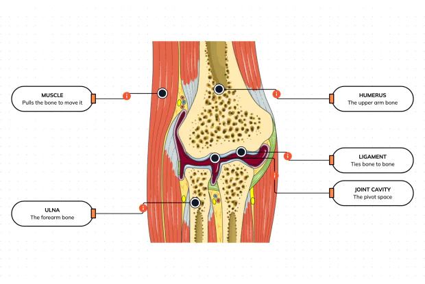
What Makes Your Elbow Bend?
Bones and joints are just the hardware; nothing moves until a muscle pulls. Label the elbow to see the parts that turn a joint into movement — the muscle that pulls, the ligament that holds bone to bone, the two bones it links, and the space they pivot in — then build it yourself in an interactive playground.
What Holds Your Elbow Together?
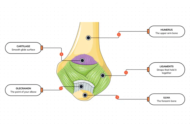
Your elbow is pulled on all day — by gravity, by every bag you carry, by every time you hang or push. So what stops the two bones from simply coming apart? Look at the back of the elbow and label the ligaments, the bony point, the cartilage and the two bones — and learn what holds the whole joint together.
What Are the Main Bones of the Skull?
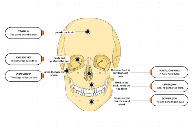
The skull looks like a single bone, but it is really about twenty-two locked together — with just one left free to move. Learn what each main part does, then label a skull yourself in an interactive playground.
What Are the Parts of Your Ear?
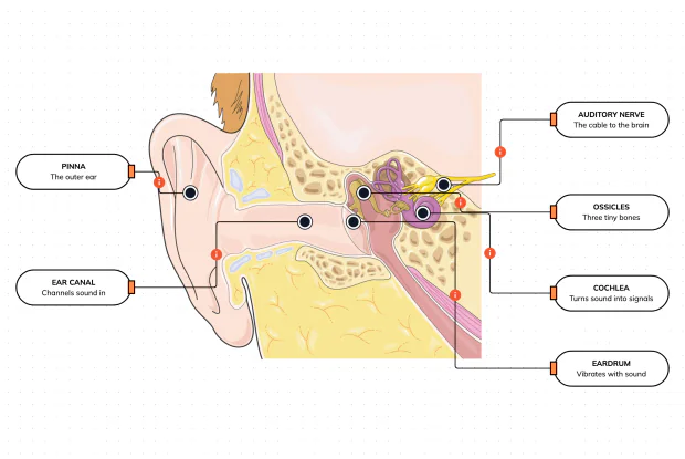
Hearing is a chain of parts passing a sound along, each handing it to the next. Label the pinna, ear canal, eardrum, ossicles, cochlea and auditory nerve, and follow a sound all the way from the air outside to a signal your brain can read — in an interactive playground.
How Does Your Ear Keep You Balanced?
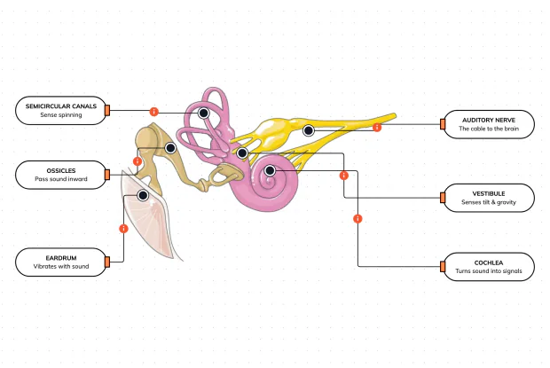
Your ear does a second, hidden job: balance. Inside it sit fluid-filled loops that sense spinning and a chamber that senses tilt and gravity. Label the semicircular canals, vestibule, cochlea, ossicles, eardrum and nerve, and learn how one organ both hears and keeps you upright — in an interactive playground.
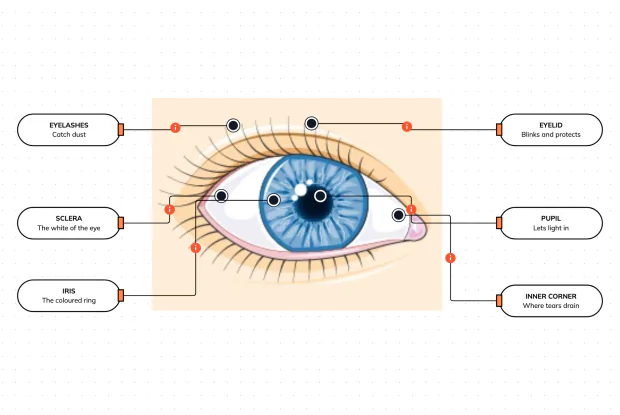
What Are the Parts of the Eye You Can See?
Before you look inside the eye, learn the parts you can already see in a mirror. Label the pupil, iris, sclera, eyelid, eyelashes and the tear-duct corner, and learn what each visible part does to let in light and protect the eye — in an interactive playground.
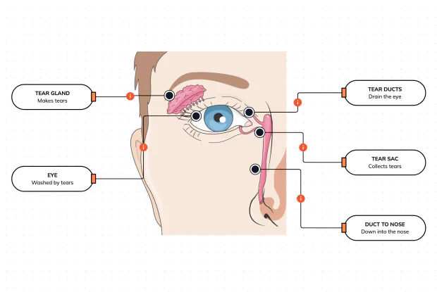
Where Do Your Tears Come From and Go?
Your eyes make and drain tears all day to stay clean and wet. Label the tear gland, the eye, the tear ducts, the tear sac and the duct to the nose, and follow a tear from where it is made to where it drains — in an interactive playground.
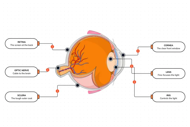
How Does Your Eye Turn Light Into a Picture?
Your eye works like a living camera. Cut it open and label the cornea, lens, iris, retina, optic nerve and sclera, and follow light as it is focused onto a screen at the back of the eye and sent to the brain — in an interactive playground.