What Are the Parts of the Femur?
Root Concept
The femur is the thigh bone — the longest and strongest bone in the body. A rounded head fits into the hip socket, a narrow neck links it to the shaft, a large greater trochanter gives hip muscles a grip, the long shaft carries the body's weight, and two rounded condyles at the bottom form the top of the knee.
CodePLU Goal
Upgrading Human Mental Models
Learn how to think in Workflows
Concept Development By codeplu.com
The five landmarks of the thigh bone, each joined to the job it does — from the ball at the hip to the condyles at the knee
Why Is the Femur the Biggest Bone You Own?
Put your hand on the front of your thigh and press. Under the muscle is a single, long, solid bone running from your hip all the way to your knee — the femur, or thigh bone. It is the longest bone in your body (roughly a quarter of your height), the strongest, and the heaviest, and it has to be: every time you stand, walk, run or jump, this one bone carries your entire body weight and passes it safely down to your knee. A bone doing that much work is not a plain rod — it is shaped, with a distinct part for each job.
Look at the two ends. At the top, a smooth round ball — the head — sits on a narrow neck and drops into a socket in your hip, making the ball-and-socket joint that lets your leg swing forwards, backwards and out to the side. Just beside the neck is a big bump, the greater trochanter, that sticks out at the side of your hip where you can actually feel it; this is where the powerful muscles of your hip and buttock grab hold to move the leg.
Between the ends runs the shaft — the long, thick, slightly curved column built to bear loads without snapping. And at the bottom the bone flares into two rounded knuckles, the condyles, capped with smooth cartilage, which meet the shin bone to form the knee. In the playground above you will label all five — the head, neck, greater trochanter, shaft and condyles — and the label on each connection tells you the job that part does.
What Does Each Part of the Femur Do?
What is the head of the femur, and why is it a ball?
The head is the smooth, rounded ball at the top of the femur, and its shape is the whole point: it drops neatly into a cup-shaped socket in your hip bone to make a ball-and-socket joint. That is the freest-moving kind of joint in the body — a ball can turn in its socket in almost any direction, which is why you can swing your leg forwards to walk, backwards, out to the side, and even rotate it. Like the other moving surface of a joint, the head is capped with smooth cartilage (drawn blue here) so it glides in the socket without grinding. A shoulder works the same way, which is why hips and shoulders are the two joints that can circle right around.
Why is the neck the femur's weak spot?
The neck is the short, narrow bridge of bone that connects the round head to the top of the shaft, and it does something clever: it holds the head off to the side at an angle, so the ball sits in the hip socket while the long shaft runs down closer to the body's centre line. That angle is what lets your feet stay under you instead of out wide. But the neck is also the thinnest part of the top of the bone, and it carries a lot of leverage, so it is the classic place the femur breaks. When an older person 'breaks a hip', it is almost always the neck of the femur that has snapped — the bone did not fail in the thick shaft, but in this narrow, highly-stressed bridge.
What is the greater trochanter — the bump you can feel?
The greater trochanter is the large bump of bone that sticks out at the top of the femur, just to the side of the neck. Rest your hand on the widest point of your hip and press — that hard lump you feel through the skin is the greater trochanter. It is not part of any joint; its job is to be an anchor. The big, powerful muscles of your hip and buttock (the ones that straighten your hip and hold you upright as you walk) attach to this bump, and because it sticks out to the side it gives those muscles leverage — like grabbing a door far from its hinge instead of close to it — so a smaller pull moves the heavy leg.
Why is the shaft built like a strong tube?
The shaft is the long, straight-ish column that makes up most of the bone's length, and it is the part that actually carries your weight from hip to knee. It looks solid, but the shaft is really a thick tube: dense, heavy compact bone on the outside wrapped around a hollow middle. That tube shape is the same trick used in scaffolding poles and bicycle frames — a tube is almost as strong as a solid rod against bending but far lighter, so the femur can be immensely strong without being impossibly heavy to lift with every stride. The shaft is also very slightly curved and bowed, which helps it flex a tiny amount under load instead of shattering, the way a springy branch bends where a dry stick snaps.
What are the condyles at the bottom?
At the knee end the femur widens into two big rounded knuckles called the condyles — a medial one on the inner side and a lateral one on the outer. They are capped with smooth cartilage (blue in the figure) and they rest on the flat top of the shin bone (the tibia) to form the knee joint. Because the condyles are rounded, they roll and glide on the shin as you bend and straighten your knee, rather than pivoting on a single point. The rounded twin-knuckle shape also makes the knee a hinge that is steady from side to side while still bending smoothly — which is exactly what you need in a joint that has to lock straight to stand yet fold to sit.
Real World Example
How Can One Bone Hold Up Your Whole Body — Yet Break at the Hip?
It sounds like a contradiction: the femur is the strongest bone you have, able to take huge forces every time you run or jump, and yet a 'broken hip' is one of the most common serious injuries in older age. Looking at the shape of the bone explains both facts at once — where it is strong, and where it is not:
The column built to bear loads
The shaft
The long shaft of the femur is a marvel of natural engineering. It is a thick tube of dense compact bone, and a tube resists bending almost as well as a solid rod while weighing far less — the same reason bikes and scaffolding are built from tubes, not bars. Its slight forward curve lets it give a little under load rather than snapping, like a springy branch. Because of this design the shaft can carry several times your body weight when you land from a jump, and it very rarely breaks in ordinary life; it takes something violent, like a car crash, to snap a healthy femoral shaft. So the middle of the bone, the part doing the obvious weight-carrying, is almost never the part that fails.
The angled bridge that takes the strain
The neck
The weakness is at the top. To place the ball into the hip socket while keeping the shaft under the body, the femur turns a corner at the neck — a short, narrow bridge of bone set at an angle. That angle is useful for walking, but it means your body weight does not press straight down the neck; it presses across it, trying to bend it, and a bar is far easier to snap sideways than to crush end-on. The neck is also thinner than the shaft. So this small bridge quietly carries the most awkward, most concentrated stress in the whole bone, every single step — which is exactly why, when the femur does break near the hip, it is the neck that gives way.
Why older people break a hip
A neck fracture
Put those two facts together and 'broken hip' makes sense. With age, bones lose density and become more brittle — and the thin, highly-stressed neck of the femur has the least bone to spare, so it is the first place to reach breaking point. A minor stumble or a fall from standing, harmless to a young skeleton, can be enough to snap the weakened neck; sometimes the neck even cracks first and causes the fall. This is why doctors watch bone density in older people and why a broken hip almost always means the femoral neck, not the mighty shaft. Understanding the bone's shape turns a confusing fact into an obvious one: the femur fails where it is thinnest and most strained, not where it looks strongest.
FAQs
Final Words
The femur is not just a rod of bone — it is a shaped tool, with a distinct part for every job. The rounded head makes a ball-and-socket joint at the hip, the angled neck bridges the head to the shaft, the greater trochanter gives the hip muscles a grip, the long tubular shaft carries your body weight, and the twin condyles form the knee. Name those five and you understand how one bone can be free-moving at the top, immensely strong in the middle, and hinge smoothly at the bottom.
Knowing the shape also explains the famous fact that a 'broken hip' happens at the femur's neck — the thinnest, most strained part — not in the mighty shaft. Every long bone in your body follows the same idea: a rounded end for the joint, a strong shaft in between, and bumps where muscles pull. Learn it once on the femur, the biggest and clearest of them all, and you can read every other bone in the skeleton.
Continue This Track
This concept is part 21 of The Human Body, Head to Toe.
What Is a Bone Made Of?
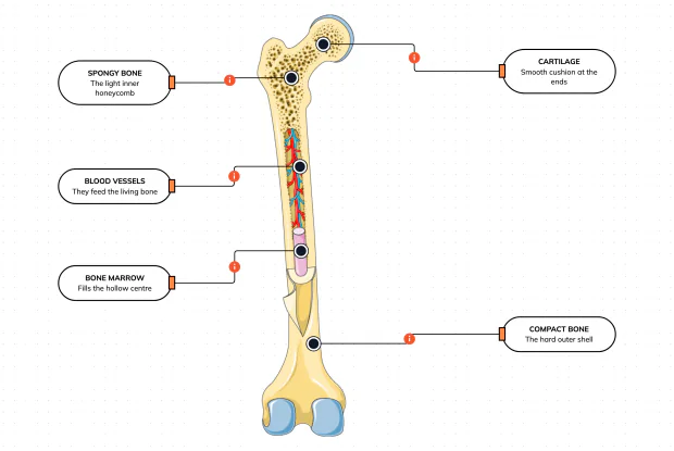
Cut a bone open and it is nowhere near solid: a hard outer shell, a light honeycomb inside, a hollow centre full of marrow, blood vessels threading through, and a smooth cushion at each end. Learn what each part does — then label them yourself in an interactive playground.
What Is Inside Your Elbow Joint?
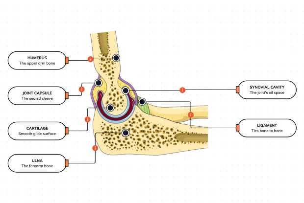
A joint is where two bones meet and move — and it is far more than just the bones. Cut open the elbow and you find smooth cartilage caps, a sealed capsule of slippery fluid, and tough ligaments holding it all together. Learn what each part does, then label them yourself in an interactive playground.
What Makes Your Elbow Bend?
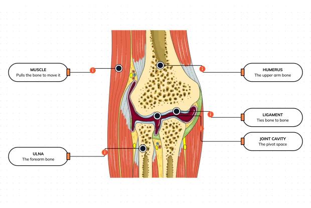
Bones and joints are just the hardware; nothing moves until a muscle pulls. Label the elbow to see the parts that turn a joint into movement — the muscle that pulls, the ligament that holds bone to bone, the two bones it links, and the space they pivot in — then build it yourself in an interactive playground.
What Holds Your Elbow Together?
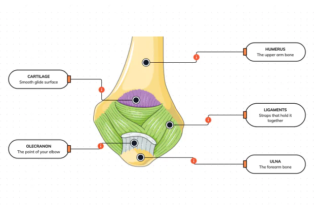
Your elbow is pulled on all day — by gravity, by every bag you carry, by every time you hang or push. So what stops the two bones from simply coming apart? Look at the back of the elbow and label the ligaments, the bony point, the cartilage and the two bones — and learn what holds the whole joint together.
What Are the Main Bones of the Skull?
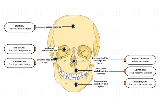
The skull looks like a single bone, but it is really about twenty-two locked together — with just one left free to move. Learn what each main part does, then label a skull yourself in an interactive playground.
What Are the Parts of Your Ear?
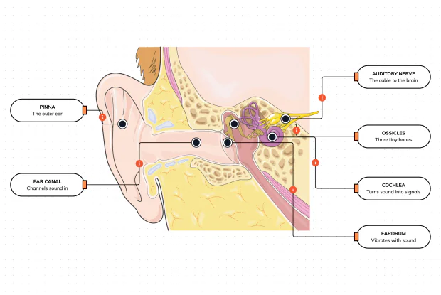
Hearing is a chain of parts passing a sound along, each handing it to the next. Label the pinna, ear canal, eardrum, ossicles, cochlea and auditory nerve, and follow a sound all the way from the air outside to a signal your brain can read — in an interactive playground.
How Does Your Ear Keep You Balanced?
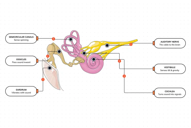
Your ear does a second, hidden job: balance. Inside it sit fluid-filled loops that sense spinning and a chamber that senses tilt and gravity. Label the semicircular canals, vestibule, cochlea, ossicles, eardrum and nerve, and learn how one organ both hears and keeps you upright — in an interactive playground.
What Are the Parts of the Eye You Can See?
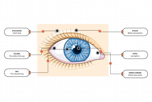
Before you look inside the eye, learn the parts you can already see in a mirror. Label the pupil, iris, sclera, eyelid, eyelashes and the tear-duct corner, and learn what each visible part does to let in light and protect the eye — in an interactive playground.
Where Do Your Tears Come From and Go?
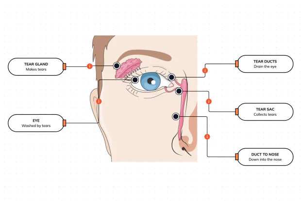
Your eyes make and drain tears all day to stay clean and wet. Label the tear gland, the eye, the tear ducts, the tear sac and the duct to the nose, and follow a tear from where it is made to where it drains — in an interactive playground.
How Does Your Eye Turn Light Into a Picture?
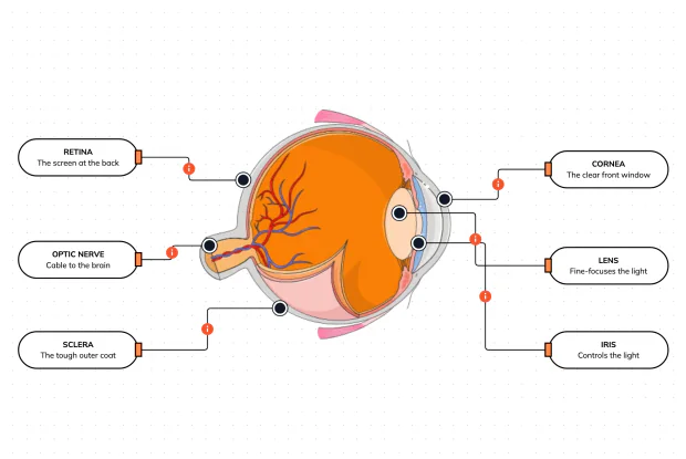
Your eye works like a living camera. Cut it open and label the cornea, lens, iris, retina, optic nerve and sclera, and follow light as it is focused onto a screen at the back of the eye and sent to the brain — in an interactive playground.