What Are the Parts of a Tooth?
Root Concept
A tooth has a hard enamel outer coat over a tough dentin layer that makes up most of it, a soft living pulp of nerves and blood vessels at its centre, and roots anchored in the jawbone, sealed at the gum, fed by a nerve and vessels entering through the root tip.
CodePLU Goal
Upgrading Human Mental Models
Learn how to think in Workflows

Concept Development By codeplu.com
A tooth in cross-section, each part joined to the job it does — from the hard enamel armour to the living pulp and anchoring roots
What Is a Tooth Actually Made Of?
A tooth looks like a simple hard white lump, but cut one in half and it turns out to be a layered structure — part armour, part living tissue — anchored deep in your jaw. Only about the top third of it, the crown, shows above the gum; the rest is roots, hidden below, holding the tooth firmly in place. Understanding those layers is what explains everything from why enamel matters to why a deep cavity is so painful.
From the outside in, there are three layers. The outermost is enamel, the hard, glossy white coat that covers the crown and does the actual biting and chewing — it is the hardest substance your body makes. Beneath it is dentin, a tough, slightly yellow layer that forms the bulk of the tooth. And right in the middle is the pulp, a soft core of living tissue packed with nerves and blood vessels. Hard on the outside, alive on the inside.
Holding it all in place is the anchoring system. The roots of the tooth sit in sockets in the jawbone, the pink gum wraps around the neck of the tooth and seals the join, and a nerve and blood vessels thread in through a tiny hole at the tip of each root to keep the pulp alive. In the playground you will label the enamel, dentin, pulp, gum, jawbone and the nerve — and the label on each connection tells you the job that part does.
What Does Each Part of a Tooth Do?
What is enamel, and why is it so hard?
Enamel is the white outer coat of the crown, and it is the hardest material your body produces — harder even than bone. It has to be, because it takes the full force of biting and chewing many times a day for a lifetime, and it shields the softer, sensitive layers underneath from hot, cold, sweet and acidic foods. Enamel gets its toughness from being made almost entirely of tightly packed mineral, with very little living material in it. That hardness is a great strength but also its one weakness: because enamel is not really 'alive', it cannot heal or grow back once it is worn away or eaten into by decay. Bone can mend a break; enamel cannot repair a hole. That single fact is why brushing, and avoiding constant sugar and acid, matters so much — you are protecting a layer that, once lost, is gone for good.
What is dentin?
Dentin is the layer just beneath the enamel, and it makes up most of the tooth — both the bulk of the crown under the white coat, and nearly all of the roots. It is slightly yellow, and it is what gives teeth their natural off-white colour showing faintly through the enamel. Dentin is hard, but not as hard as enamel, and, importantly, it is partly living: it is threaded with millions of microscopic tubes that connect to the nerves in the pulp. This is why dentin can feel things. When a cavity eats through the enamel and reaches the dentin, you start to notice sensitivity — a twinge with something cold or sweet — because those tiny tubes carry the signal inward. Dentin is the tooth's tough middle layer, softer and more alive than the enamel guarding it, and it is the bridge between the dead-hard surface and the living core.
What is the pulp?
The pulp is the soft tissue at the very centre of the tooth — the part that is fully alive. It fills a chamber in the crown and runs down narrow canals inside each root, and it is packed with nerves and tiny blood vessels. The blood vessels keep the tooth nourished, and the nerves are what let a tooth feel: pressure, temperature, and, unfortunately, pain. The pulp is the reason a tooth is a living organ and not just a hard peg. It also explains toothache: because the pulp is where the nerves are, anything that reaches or inflames it — deep decay, a crack, an infection — hurts intensely. When a dentist does a 'root canal', they are removing damaged pulp from those inner canals. Tucked safely in the middle, shielded by dentin and enamel, the pulp is the living heart of the tooth.
How is a tooth held in the jaw?
A tooth is not simply stuck onto the jaw — its roots are set into deep sockets in the jawbone, like tent pegs driven into the ground, which is what lets a tooth withstand the strong forces of chewing without wobbling loose. The roots are the lower part of the tooth, below the crown, and a large tooth like a molar has two or three of them for extra grip. Wrapping around the neck of the tooth, where crown meets root, is the gum — the firm pink tissue that hugs the tooth and seals the join, keeping food and bacteria from getting down to the roots and bone. Healthy gum is essential: if it becomes diseased and pulls away, the anchoring is weakened and teeth can loosen. So the jawbone provides the anchor, and the gum provides the seal.
What keeps a tooth alive?
A living tooth needs a blood supply and a nerve, and both reach it the same way: through a tiny opening at the very tip of each root. A small bundle of nerve and blood vessels runs up from the jaw, enters that hole, and travels through the root canal to reach the pulp in the centre. The blood vessels bring oxygen and nutrients to keep the pulp healthy, and the nerve carries sensation back out. This is why a tooth can 'die' — if that supply is cut off or the pulp becomes badly infected, the living tissue inside dies even though the hard shell remains. It is also why deep decay is serious: once bacteria reach the pulp and its nerve, the pain is severe and the tooth's life is at risk. That slender bundle entering the root tip is the tooth's lifeline to the rest of the body.
Real World Example
Why Does a Cavity Start Painless but End Agonising?
A cavity is bacteria slowly eating a hole from the outside in, and the tooth's layered structure explains exactly why it hurts when it does:
Through the armour, unnoticed
Decay in the enamel
Decay begins on the enamel, the hard outer coat, where bacteria feeding on sugar produce acid that slowly dissolves the mineral. At this stage you feel nothing at all, because enamel has no nerves — it is essentially non-living armour. This is what makes early decay so dangerous: it can be quietly eating into a tooth for a long time with no warning sign, which is precisely why dentists check for it and why brushing is preventive rather than a response to pain. And because enamel cannot repair itself, any hole the acid makes is permanent and will only get bigger unless a dentist cleans it out and fills it. The painless start is not good news — it is the window in which the damage is easiest to stop but hardest to notice.
Into the dentin, the first twinges
Decay reaching the dentin
Once the decay breaks through the enamel and reaches the dentin, you start to feel it. Dentin is softer, so the cavity now grows faster, and — crucially — it is threaded with tiny tubes leading to the nerves in the pulp. Cold, hot and sweet things can now travel down those tubes and produce a sharp twinge, the classic sign of a cavity that has got past the surface. This is the stage where most people finally notice something is wrong. The tooth is warning you that the decay has left the dead outer shell and entered a living, connected layer. Acting now, with a filling, stops the decay before it reaches the most sensitive part — the difference between a simple repair and a serious problem.
Into the pulp, full toothache
Decay reaching the pulp
If the decay is left to continue, it eventually reaches the pulp — the living core full of nerves and blood vessels — and now the pain is severe and constant, the throbbing toothache everyone dreads. Bacteria in the pulp cause it to become inflamed and infected in a tight space it cannot escape, pressing on the nerve. At this point a simple filling is no longer enough: the dentist usually has to remove the damaged pulp entirely in a root canal, or, if it is too far gone, take the tooth out. This final stage is the whole reason the earlier ones matter: the pain is the tooth telling you the decay has finally reached the part that is alive. The journey from painless surface to agonising centre is a journey through the very layers you have just labelled.
FAQs
Final Words
A tooth is a layered structure: hard enamel armour on the outside, tough dentin making up most of the body, and a soft living pulp of nerves and blood vessels at the centre. Its roots are anchored in sockets in the jawbone, the gum seals the join, and a nerve and vessels enter through the root tip to keep it alive. Hard on the outside, alive on the inside, and firmly pegged into the jaw.
Those layers explain the whole story of tooth decay — painless while it eats through the dead enamel, twinging once it reaches the living dentin, and agonising when it finally reaches the pulp. It is a perfect example of how knowing the parts of something explains what happens to it. This tooth is where digestion begins, doing the chewing; next in the mouth comes the tongue, which moves the food and lets you taste it.
Continue This Track
This concept is part 25 of The Human Body, Head to Toe.
What Is a Bone Made Of?
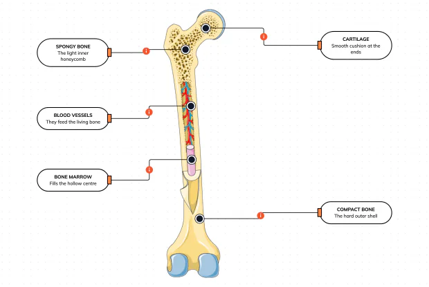
Cut a bone open and it is nowhere near solid: a hard outer shell, a light honeycomb inside, a hollow centre full of marrow, blood vessels threading through, and a smooth cushion at each end. Learn what each part does — then label them yourself in an interactive playground.
What Is Inside Your Elbow Joint?
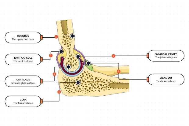
A joint is where two bones meet and move — and it is far more than just the bones. Cut open the elbow and you find smooth cartilage caps, a sealed capsule of slippery fluid, and tough ligaments holding it all together. Learn what each part does, then label them yourself in an interactive playground.
What Makes Your Elbow Bend?
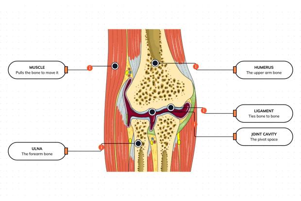
Bones and joints are just the hardware; nothing moves until a muscle pulls. Label the elbow to see the parts that turn a joint into movement — the muscle that pulls, the ligament that holds bone to bone, the two bones it links, and the space they pivot in — then build it yourself in an interactive playground.
What Holds Your Elbow Together?
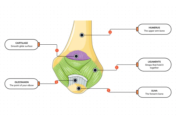
Your elbow is pulled on all day — by gravity, by every bag you carry, by every time you hang or push. So what stops the two bones from simply coming apart? Look at the back of the elbow and label the ligaments, the bony point, the cartilage and the two bones — and learn what holds the whole joint together.
What Are the Main Bones of the Skull?
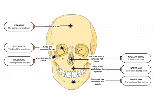
The skull looks like a single bone, but it is really about twenty-two locked together — with just one left free to move. Learn what each main part does, then label a skull yourself in an interactive playground.
What Are the Parts of Your Ear?
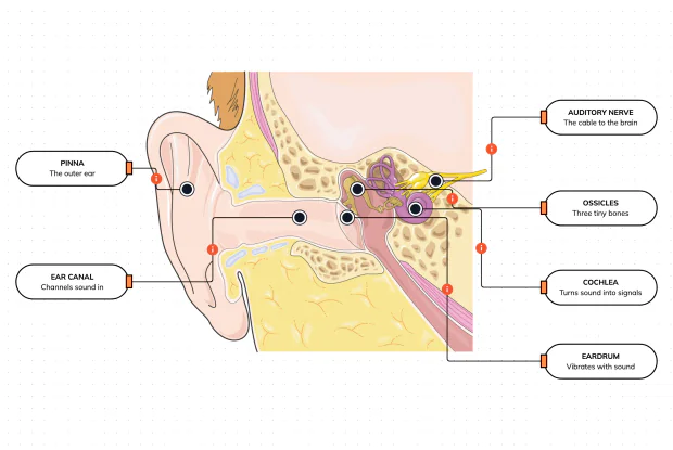
Hearing is a chain of parts passing a sound along, each handing it to the next. Label the pinna, ear canal, eardrum, ossicles, cochlea and auditory nerve, and follow a sound all the way from the air outside to a signal your brain can read — in an interactive playground.
How Does Your Ear Keep You Balanced?
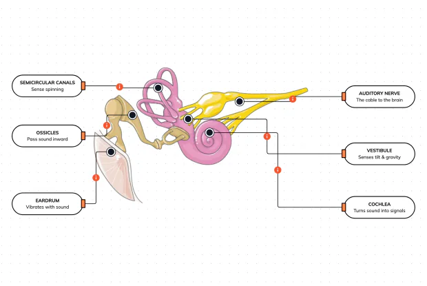
Your ear does a second, hidden job: balance. Inside it sit fluid-filled loops that sense spinning and a chamber that senses tilt and gravity. Label the semicircular canals, vestibule, cochlea, ossicles, eardrum and nerve, and learn how one organ both hears and keeps you upright — in an interactive playground.
What Are the Parts of the Eye You Can See?
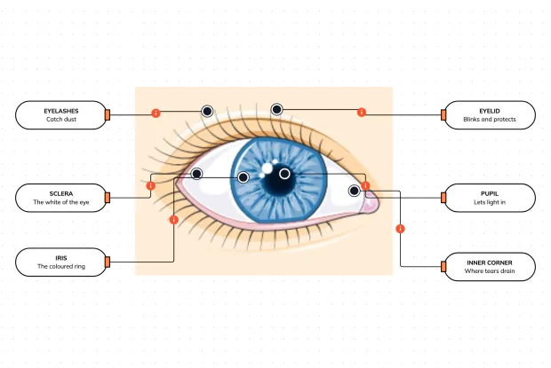
Before you look inside the eye, learn the parts you can already see in a mirror. Label the pupil, iris, sclera, eyelid, eyelashes and the tear-duct corner, and learn what each visible part does to let in light and protect the eye — in an interactive playground.
Where Do Your Tears Come From and Go?
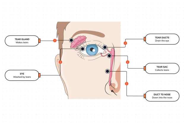
Your eyes make and drain tears all day to stay clean and wet. Label the tear gland, the eye, the tear ducts, the tear sac and the duct to the nose, and follow a tear from where it is made to where it drains — in an interactive playground.
How Does Your Eye Turn Light Into a Picture?
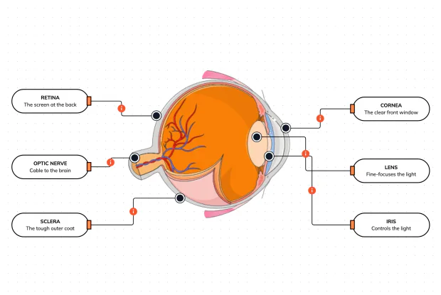
Your eye works like a living camera. Cut it open and label the cornea, lens, iris, retina, optic nerve and sclera, and follow light as it is focused onto a screen at the back of the eye and sent to the brain — in an interactive playground.