How Does the Pancreas Deliver Its Juice?
Root Concept
The pancreas collects its digestive juice through a branching duct system: small branches gather juice from all over the gland into a main pancreatic duct that runs from the tail toward the head. Near the head it joins the bile duct from the liver, and the two empty together into the small intestine through a shared opening.
CodePLU Goal
Upgrading Human Mental Models
Learn how to think in Workflows

Concept Development By codeplu.com
The pancreas's duct system, each part joined to the job it does — collecting digestive juice and piping it to the gut, where it meets the bile duct
How Does the Pancreas Get Its Juice to Your Gut?
The pancreas makes a powerful digestive juice throughout its whole length — but making it is only half the story. That juice is useless sitting inside the gland; it has to be delivered to the small intestine, where the food is, and delivered on demand when a meal arrives. This view cuts the pancreas open to show the pipework that does exactly that: a branching network of ducts that gathers the juice from every corner of the gland and channels it to a single exit.
The system works just like a river and its tributaries, only in reverse. All across the gland, tiny amounts of juice are made and emptied into small duct branches. These branches join into larger ones, which all feed into one main pancreatic duct running the length of the organ, from the thin tail at one end toward the wide head at the other. As it travels, the main duct collects more and more juice from each branch it passes, so by the time it reaches the head it is carrying the output of the entire gland.
At the head, something important happens: the pancreatic duct meets the bile duct coming down from the liver. Instead of each having its own separate exit, the two usually join and empty together through a single shared opening into the small intestine. This shared exit is elegant — pancreatic juice and bile arrive at the food together — but it also creates a vulnerability, because one blockage there can stop both. In the playground above you will label the main duct, a branch, the bile duct, the head and the tail — and the label on each connection tells you the job that part does.
How Does the Pancreatic Duct System Work?
What is the pancreatic duct?
The pancreatic duct is the main channel that carries the pancreas's digestive juice out of the gland and toward the gut. It runs almost the entire length of the pancreas, from the tail at the thin end to the head at the wide end, like a central pipe threaded through the organ. It does not make anything itself; its job is purely transport — to collect the juice produced all along the gland and deliver it to where it is needed, the small intestine. You can see it in this cut-open view as the prominent tube running lengthwise through the pale tissue. Everything the pancreas produces for digestion travels down this one duct, which makes it the gland's essential plumbing.
Why does the duct have so many branches?
Because the juice is made everywhere in the gland, not in one spot — so it has to be collected from everywhere. The pancreas is packed with millions of tiny juice-making clusters spread throughout its whole body, and each empties into a small duct nearby. These little ducts join into slightly bigger branches, which join into bigger ones still, all eventually feeding the single main duct. The pattern is exactly like the veins of a leaf or the tributaries of a river: many small streams gathering into one main channel. This branching design is efficient — it lets the gland collect juice from its entire volume without any part being too far from a duct — and it is why the main duct, cut open here, has a feathery tree of branches feeding into it along its length.
Why does the bile duct join the pancreatic duct?
Near the head of the pancreas, the pancreatic duct is met by the bile duct — the tube bringing bile down from the liver and gallbladder. Rather than each punching its own separate hole into the intestine, the two ducts usually merge and share a single exit. This makes good sense: bile and pancreatic juice do complementary digestive jobs — bile breaks fat into droplets, and the pancreatic enzymes then digest that fat, along with proteins and carbohydrates — so delivering them to the food at the same place and the same time is efficient. It is a bit like two supply lines meeting at one loading dock right where the goods are needed. The green bile duct entering near the head in this figure shows that meeting point.
Where do the juices empty?
Both juices empty into the duodenum, the first part of the small intestine, through the shared opening at the head of the pancreas. This is exactly where the head is positioned for — tucked into the curve of the duodenum so its outlet sits right against the intestine. The opening is guarded by a small ring of muscle (a sphincter) that acts as a valve: it stays closed most of the time and opens to release bile and pancreatic juice when a meal arrives and needs digesting. So the delivery is not a constant dribble but a controlled release, timed to the food. This is why the pancreatic and bile systems are so closely tied to eating — their whole output is aimed at that one guarded doorway into the gut.
What do the head and tail tell you about the flow?
The head and tail mark the two ends of the duct's journey, and knowing them tells you which way the juice flows. The tail is the thin far end of the pancreas, over near the spleen, and it is where the main duct begins as little more than a trickle. From there the duct runs toward the head, the wide end tucked into the intestine, gathering juice from branch after branch as it goes — so the flow is always tail-to-head, ending at the exit into the gut. The head is therefore the busy end: not only the outlet for the whole gland's juice, but also the junction with the bile duct. That is why problems at the head — a blockage or a tumour — are so serious: they sit right at the single point everything has to pass through.
Real World Example
Why Can One Small Gallstone Cause Two Big Problems at Once?
The pancreas's duct system is a model of efficient plumbing, but the shared exit with the bile duct is a double-edged design. Three features show why — and where it can go wrong:
A river and its tributaries
The branching duct
The pancreas faces a collection problem: it makes juice throughout its whole volume, so it needs to gather that juice from everywhere and funnel it to one exit. Its solution is the same one nature uses for draining a landscape — a branching tree of ducts, like a river system. Tiny ducts collect juice locally, join into larger branches, and finally merge into the single main duct, which carries the combined output to the head. This design means no part of the gland is far from drainage, and the whole gland's juice can be delivered through one controlled outlet. It is the same efficient pattern you see in the veins of a leaf, the airways of the lungs, and the blood vessels of the body: many small channels gathering into a few large ones.
Two ducts, one exit
The shared junction with the bile duct
Near the head, the pancreatic duct and the bile duct usually join and empty through one shared opening into the intestine. This is efficient — bile and pancreatic juice work on food together, so arriving at the same place and time is ideal, and the body needs to guard only one doorway with one valve instead of two. For almost everyone, almost all the time, this shared exit works flawlessly, releasing both juices in a coordinated burst whenever a meal arrives. It is a neat piece of biological engineering: combine two supply lines at the last moment so they reach their destination together. But merging two systems into one exit means they now share a single point of failure — which is where trouble can start.
When a stone blocks the shared exit
Gallstone pancreatitis
Here is the danger of the shared design. Gallstones form in the gallbladder and can travel down the bile duct; usually the worst they do is block bile and cause jaundice. But if a stone lodges right at the shared opening — below where the pancreatic duct has already joined — it blocks not just the bile but the pancreatic duct too. Now the pancreas's powerful juice cannot escape, backs up inside the gland, and its enzymes can switch on where they should not, so the pancreas begins to digest itself. This is gallstone pancreatitis, a sudden and severe illness, and it happens precisely because the two ducts share one exit. One small stone, in one unlucky spot, jams two systems at once — a vivid lesson in how the same shared plumbing that makes the pancreas efficient also makes it vulnerable.
FAQs
Final Words
Making digestive juice is only half the pancreas's job — it also has to deliver it, and this cut-open view shows the pipework that does. A branching network of ducts gathers juice from every part of the gland, like tributaries joining a river, into one main pancreatic duct that runs from the tail toward the head. There it meets the bile duct from the liver, and the two empty together through a single guarded opening into the small intestine, releasing their juices when a meal arrives.
That shared exit is a neat piece of design — two complementary juices delivered to the food at once — but it comes with a catch: one blockage can stop both, which is why a gallstone in the wrong place can trigger pancreatitis. Seeing the pancreas as an organ, and now as a delivery system, completes the picture of how it feeds your digestion. The organ-systems tour of the body carries on from here.
Continue This Track
This concept is part 33 of The Human Body, Head to Toe.
What Is a Bone Made Of?
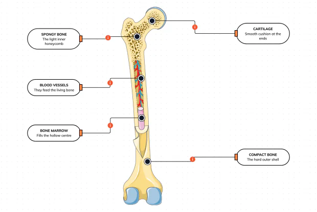
Cut a bone open and it is nowhere near solid: a hard outer shell, a light honeycomb inside, a hollow centre full of marrow, blood vessels threading through, and a smooth cushion at each end. Learn what each part does — then label them yourself in an interactive playground.
What Is Inside Your Elbow Joint?
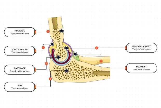
A joint is where two bones meet and move — and it is far more than just the bones. Cut open the elbow and you find smooth cartilage caps, a sealed capsule of slippery fluid, and tough ligaments holding it all together. Learn what each part does, then label them yourself in an interactive playground.
What Makes Your Elbow Bend?
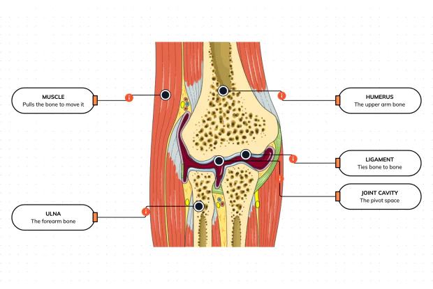
Bones and joints are just the hardware; nothing moves until a muscle pulls. Label the elbow to see the parts that turn a joint into movement — the muscle that pulls, the ligament that holds bone to bone, the two bones it links, and the space they pivot in — then build it yourself in an interactive playground.
What Holds Your Elbow Together?
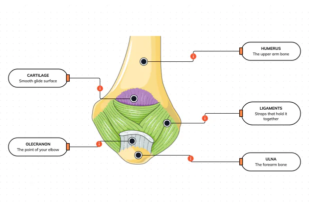
Your elbow is pulled on all day — by gravity, by every bag you carry, by every time you hang or push. So what stops the two bones from simply coming apart? Look at the back of the elbow and label the ligaments, the bony point, the cartilage and the two bones — and learn what holds the whole joint together.
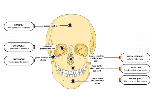
What Are the Main Bones of the Skull?
The skull looks like a single bone, but it is really about twenty-two locked together — with just one left free to move. Learn what each main part does, then label a skull yourself in an interactive playground.
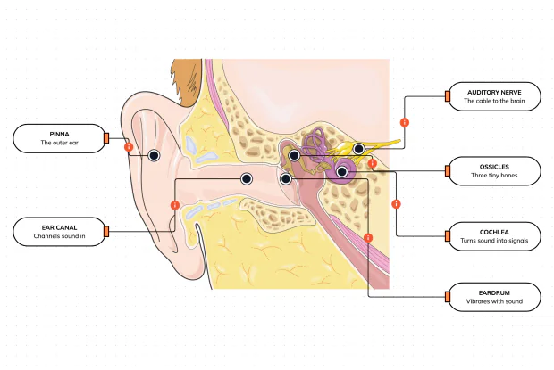
What Are the Parts of Your Ear?
Hearing is a chain of parts passing a sound along, each handing it to the next. Label the pinna, ear canal, eardrum, ossicles, cochlea and auditory nerve, and follow a sound all the way from the air outside to a signal your brain can read — in an interactive playground.
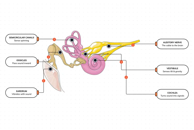
How Does Your Ear Keep You Balanced?
Your ear does a second, hidden job: balance. Inside it sit fluid-filled loops that sense spinning and a chamber that senses tilt and gravity. Label the semicircular canals, vestibule, cochlea, ossicles, eardrum and nerve, and learn how one organ both hears and keeps you upright — in an interactive playground.
What Are the Parts of the Eye You Can See?
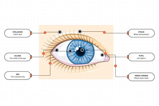
Before you look inside the eye, learn the parts you can already see in a mirror. Label the pupil, iris, sclera, eyelid, eyelashes and the tear-duct corner, and learn what each visible part does to let in light and protect the eye — in an interactive playground.
Where Do Your Tears Come From and Go?
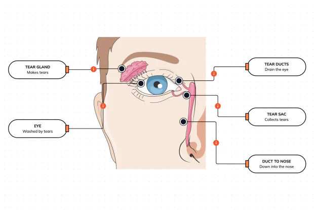
Your eyes make and drain tears all day to stay clean and wet. Label the tear gland, the eye, the tear ducts, the tear sac and the duct to the nose, and follow a tear from where it is made to where it drains — in an interactive playground.
How Does Your Eye Turn Light Into a Picture?
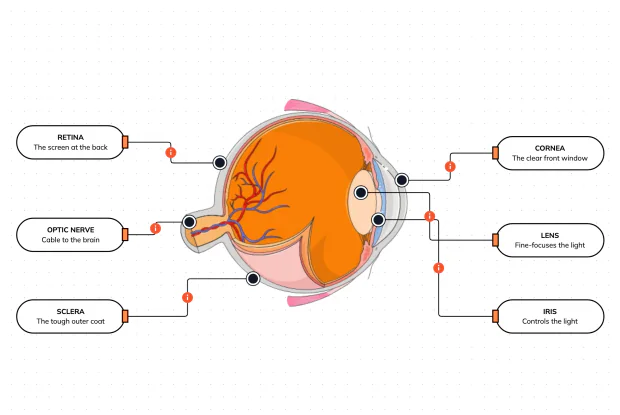
Your eye works like a living camera. Cut it open and label the cornea, lens, iris, retina, optic nerve and sclera, and follow light as it is focused onto a screen at the back of the eye and sent to the brain — in an interactive playground.