What Is an Ovarian Follicle?
Root Concept
A follicle is a ball of helper cells wrapped around one egg. The cells feed and protect the egg and make hormones, a pocket of fluid opens up inside as the follicle grows, and once the egg is released the leftover sac changes job completely and becomes a hormone-making body.
CodePLU Goal
Upgrading Human Mental Models
Learn how to think in Workflows

Concept Development By codeplu.com
A follicle close up — one egg cell, a wall of helper cells and a pocket of fluid — shown beside the smaller sizes it grows from and the yellow body it leaves behind
Why Does an Egg Need a Sac Around It?
An egg is a single cell. It is the largest cell in the human body, but it is still only one cell, and it cannot feed itself, defend itself or decide when it is ready. So it never sits alone. Every egg inside an ovary is wrapped in a ball of helper cells, and that whole package — helpers plus egg — is called a follicle.
Look at the big cut-open follicle in the playground and you can see three things at once: the egg itself, the wall of cells packed around it, and a pocket of fluid that has opened up inside. That fluid pocket is the clue that this follicle is well on its way. A follicle starts out so small it is barely more than the egg and a thin layer of cells, and it swells over weeks as the helper cells multiply and fluid collects.
The smaller shapes around the edge of the picture are the same structure at different points in its life — a part-grown one, a couple still at their starting size, and the yellow body that is left after the egg has been released. In the playground you will label the parts of a follicle and the sizes it passes through.
What Is a Follicle Made Of, and How Does It Change?
What is actually inside a follicle?
Three things, and it helps to keep them separate. First, the egg cell itself, sitting off to one side rather than in the middle — a single cell with its nucleus, the dark spot holding the genetic instructions. Second, the wall: a thick layer of helper cells wrapped all the way round. These are not spare parts. They pass food and signals inward to the egg, and they make hormones that travel outward into the blood to prepare the rest of the body. Third, the fluid, which collects in a growing pocket between the helper cells. Many beginners assume the follicle is the egg, or a shell around it. It is neither: it is a working nursery of thousands of cells, and the egg is its single occupant.
Why do follicles come in such different sizes?
Because you are seeing different moments in a slow process. A resting follicle is tiny — one egg with a thin single layer of cells around it — and it can sit like that, unchanged, for years or even decades. When one is finally woken, its helper cells begin dividing, the wall thickens, and a small pool of fluid appears and then spreads into a large space that pushes the egg to one edge. By the time a follicle is ripe it may be a couple of centimetres across, big enough to bulge from the surface of the ovary, though the egg inside has hardly changed size at all. So the growth you see is almost entirely the nursery growing, not the egg.
What happens to the sac after the egg leaves?
When a ripe follicle bursts and lets its egg out, the sac does not simply collapse and vanish. The leftover helper cells stay put, seal the gap, fill with a yellow fatty pigment, and switch to a new job: making hormones full time. That new structure is the corpus luteum, the yellow body you can see at the bottom of the picture, and its hormones keep the lining of the uterus thick and ready in case the egg is fertilised. It lasts roughly two weeks unless a pregnancy begins, then shrinks away to a small pale scar. So a follicle has two careers — first a nursery for one egg, then a temporary gland.
Real World Example
Why Doctors Look at Follicles, Not Eggs
An egg is far too small to see on an ultrasound scan. Follicles are not, and that single fact shapes how fertility is measured:
Counting the sacs
Follicles are visible, eggs are not
A human egg is about a tenth of a millimetre across — invisible on any normal scan. A ripening follicle, though, grows to a centimetre or two and is full of fluid, which shows up on ultrasound as a clear dark circle. So when a clinic assesses someone's fertility, they count and measure follicles and use them as a stand-in for eggs, since each sac contains one. This is why you may hear about a 'follicle count' rather than an egg count. The nursery is what can be seen; the occupant is taken on trust, because a follicle of the right size at the right moment almost always has an egg inside it.
Watching one get ready
Size tells you how close it is
Because a follicle swells steadily as it ripens, its diameter works like a clock. Repeat scans over several days show the leading follicle growing a couple of millimetres a day, and when it reaches roughly the size of a grape it is close to bursting. That lets a clinic predict ovulation within a day or so, which matters enormously for timing treatment. It also shows what is happening to the others: several may start, but one pulls ahead in size while the rest stop growing and fade. You can watch the monthly competition play out on the screen, measured in millimetres.
What this tells you
The sac is the working part
Almost everything the ovary does for the rest of the body is done by follicles rather than by eggs. They make the hormones that thicken the uterus lining, that drive the monthly cycle, and that cause the changes of puberty. The egg is precious but passive; the cells around it do the work, send the signals and set the timing. That is also why a follicle can carry on being useful after its egg has gone — as the corpus luteum, still making hormones. Understanding the follicle, not just the egg, is what makes the whole reproductive cycle finally make sense.
FAQs
Final Words
A follicle is the egg's nursery: one egg cell, a thick wall of helper cells that feed it and make hormones, and a pocket of fluid that opens up as the whole sac ripens. Follicles start out microscopic and can wait like that for decades, then swell over weeks until one is ripe enough to burst and release its egg. What grows is the nursery, not the egg.
The sac's story does not end there — once empty it becomes the corpus luteum and keeps working as a small hormone gland. Hold on to that idea and the monthly cycle stops being a list of names: it is one sac growing, opening and changing job. Next, step back to see all of these stages at once inside a whole ovary, or follow the released egg on its journey down the fallopian tube.
Continue This Track
This concept is part 60 of The Human Body, Head to Toe.
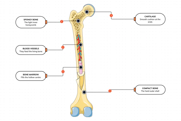
What Is a Bone Made Of?
Cut a bone open and it is nowhere near solid: a hard outer shell, a light honeycomb inside, a hollow centre full of marrow, blood vessels threading through, and a smooth cushion at each end. Learn what each part does — then label them yourself in an interactive playground.
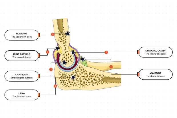
What Is Inside Your Elbow Joint?
A joint is where two bones meet and move — and it is far more than just the bones. Cut open the elbow and you find smooth cartilage caps, a sealed capsule of slippery fluid, and tough ligaments holding it all together. Learn what each part does, then label them yourself in an interactive playground.
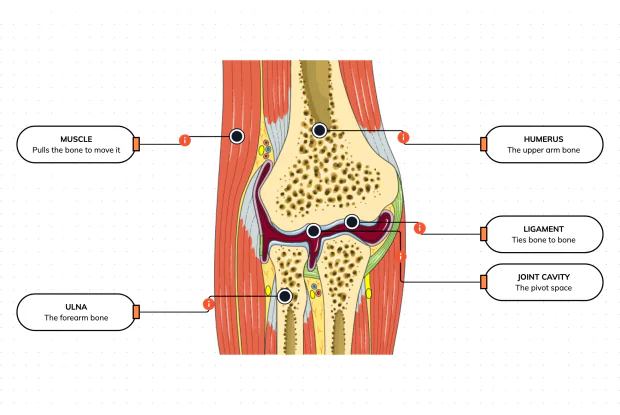
What Makes Your Elbow Bend?
Bones and joints are just the hardware; nothing moves until a muscle pulls. Label the elbow to see the parts that turn a joint into movement — the muscle that pulls, the ligament that holds bone to bone, the two bones it links, and the space they pivot in — then build it yourself in an interactive playground.
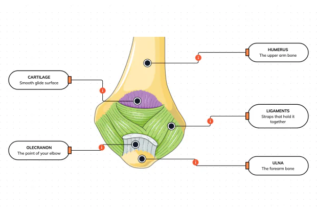
What Holds Your Elbow Together?
Your elbow is pulled on all day — by gravity, by every bag you carry, by every time you hang or push. So what stops the two bones from simply coming apart? Look at the back of the elbow and label the ligaments, the bony point, the cartilage and the two bones — and learn what holds the whole joint together.
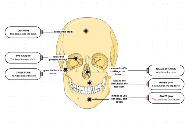
What Are the Main Bones of the Skull?
The skull looks like a single bone, but it is really about twenty-two locked together — with just one left free to move. Learn what each main part does, then label a skull yourself in an interactive playground.
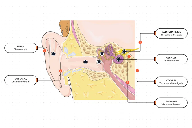
What Are the Parts of Your Ear?
Hearing is a chain of parts passing a sound along, each handing it to the next. Label the pinna, ear canal, eardrum, ossicles, cochlea and auditory nerve, and follow a sound all the way from the air outside to a signal your brain can read — in an interactive playground.
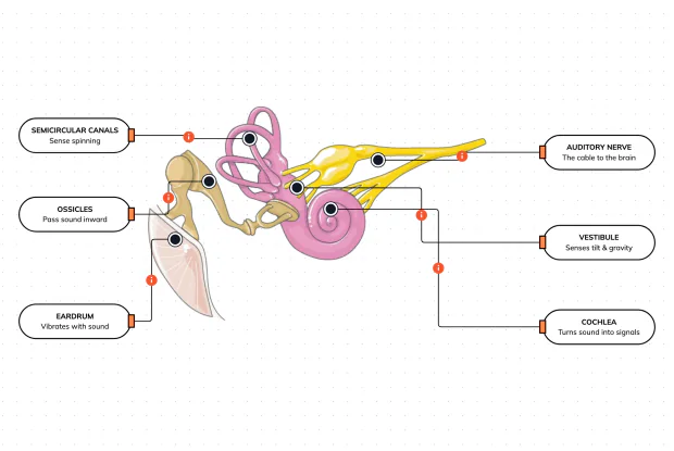
How Does Your Ear Keep You Balanced?
Your ear does a second, hidden job: balance. Inside it sit fluid-filled loops that sense spinning and a chamber that senses tilt and gravity. Label the semicircular canals, vestibule, cochlea, ossicles, eardrum and nerve, and learn how one organ both hears and keeps you upright — in an interactive playground.
What Are the Parts of the Eye You Can See?
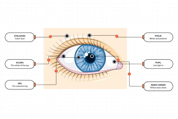
Before you look inside the eye, learn the parts you can already see in a mirror. Label the pupil, iris, sclera, eyelid, eyelashes and the tear-duct corner, and learn what each visible part does to let in light and protect the eye — in an interactive playground.
Where Do Your Tears Come From and Go?
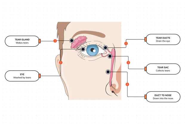
Your eyes make and drain tears all day to stay clean and wet. Label the tear gland, the eye, the tear ducts, the tear sac and the duct to the nose, and follow a tear from where it is made to where it drains — in an interactive playground.
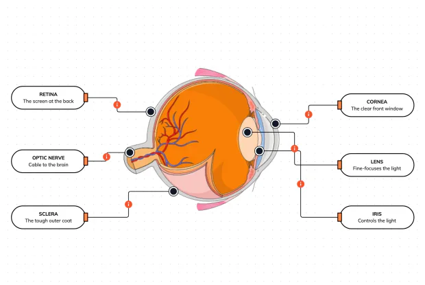
How Does Your Eye Turn Light Into a Picture?
Your eye works like a living camera. Cut it open and label the cornea, lens, iris, retina, optic nerve and sclera, and follow light as it is focused onto a screen at the back of the eye and sent to the brain — in an interactive playground.