How Do Signals Travel From Your Eyes to Your Brain?
Root Concept
The eye captures light and turns it into signals, but seeing happens in the brain. Signals leave each eye along the optic nerve, meet at the optic chiasm where the two eyes' fibres partly cross and swap sides, travel on through the optic radiations, and reach the visual cortex at the back of the brain, which builds the picture you actually see.
CodePLU Goal
Upgrading Human Mental Models
Learn how to think in Workflows

Concept Development By codeplu.com
The pathway from eyes to brain, each part joined to the job it does as a visual signal travels to where seeing happens
Where Does Seeing Actually Happen — the Eye, or the Brain?
It feels as though you see with your eyes. But your eyes are only cameras: they catch the light and turn it into electrical signals. Nothing is actually 'seen' until those signals reach the brain and the brain makes sense of them. Proof of this is simple — a perfectly healthy eye is useless if the wiring to the brain is cut, and a person can be blind with flawless eyes if the seeing part of the brain is damaged. This figure, looking up at the underside of the brain, shows that wiring: the whole route a visual signal travels from eye to brain.
The journey starts as each eye turns its image into signals and sends them down its optic nerve — the thick cable leaving the back of the eyeball. The two optic nerves head inward and meet at a spot called the optic chiasm, and something clever happens there: the fibres partly cross over, swapping sides, so that everything from the left half of your view is gathered together to go to the right side of the brain, and everything from the right half goes to the left. Your brain reorganises the two eyes' views by which side of the world they came from, not by which eye they came from.
After the crossover the signals travel on and fan out through the optic radiations — broad sweeps of fibres curving toward the back of the head — until they arrive at the visual cortex, the region at the very back of the brain where seeing truly happens. Only there are the signals finally assembled into the rich, moving, full-colour picture you experience. In the playground above you will label the eye, optic nerve, chiasm, radiation and visual cortex, and trace the signal all the way home.
How Does the Visual Signal Travel to the Brain?
If the eye captures the image, why isn't that 'seeing'?
Because the eye only does the first step. The eye focuses light onto the retina and the retina turns it into a pattern of electrical signals — but a pattern of signals is not yet an experience of seeing, any more than a photo file is a picture until something opens it. Those signals have to be carried to the brain and interpreted before you experience anything at all. This is why 'seeing' is really a partnership: the eye is the camera that captures, and the brain is where the capture becomes sight. Everything after the retina in this figure — the nerve, the crossover, the radiations, the cortex — is about getting the signal to the brain and handing it to the part that actually sees.
What does the optic nerve do?
The optic nerve is the thick cable that leaves the back of each eyeball and carries its signals toward the brain. Every one of the retina's millions of signals is bundled into this single nerve — it is the one and only route out of the eye. That also makes it a single point of failure: damage the optic nerve and the eye goes blind even though the eyeball itself is perfect, because the signals can no longer get out. There are two optic nerves, one from each eye, and in this view you can see them heading inward from the two eyeballs toward the middle, where they are about to meet and do something surprising.
What is the optic chiasm, and why do the nerves cross?
The optic chiasm is the X-shaped junction in the middle where the two optic nerves meet — and it is where the wiring is cleverly rearranged. Instead of each eye simply sending its signal to its own side of the brain, the fibres partly cross over here: the half of each eye's view that shows the left side of the world is routed to the right side of the brain, and the half showing the right side of the world goes to the left. The result is that the brain sorts vision by side of space, not by eye. Each side of your brain receives the matching half of the view from both eyes at once, which is exactly what it needs to merge the two slightly different images into a single, three-dimensional picture.
What are the optic radiations?
After the crossover, the signals continue and then spread out into the optic radiations — broad, fanned-out sweeps of nerve fibres that curve back through the brain toward the visual cortex. Think of them as the last stretch of cabling, carrying the sorted signals on their final journey to the back of the head. They fan out because the visual cortex needs the picture spread across a wide area of brain, with different fibres delivering different parts of the view to different patches of cortex. In the figure they are the wide, curving bundles sweeping toward the base of the brain. Because they are so spread out, an injury to one part of the radiations knocks out only a matching part of a person's vision rather than all of it.
What does the visual cortex do?
The visual cortex is the region at the very back of the brain (the occipital lobe) where seeing finally happens — the destination of the whole pathway. Here the incoming signals are assembled and interpreted: edges, colours, movement, depth and shapes are pieced together into the seamless, meaningful picture you actually experience. It is the visual cortex, not the eye, that recognises a face, notices motion, or reads these words. This is why damage to the back of the brain can cause blindness even when the eyes and nerves are completely healthy — the camera and cabling work, but there is nothing at the far end to turn the signal into sight. Seeing is something your brain does; the eye just supplies the raw material.
Real World Example
How Can Someone Go Blind While Their Eyes Are Perfectly Healthy?
Because vision travels this fixed route, damage at different points causes very different, very specific losses. Each one points to a part of the pathway — and to the fact that seeing happens in the brain:
Blindness from the brain, not the eye
The visual cortex
A person can have perfectly healthy eyes — clear corneas, working retinas, intact optic nerves — and still be unable to see, if the visual cortex at the back of the brain is damaged, for example by a stroke. The eyes still capture the light and send the signals faithfully, but there is nothing at the destination to turn those signals into sight, so the person is blind. In some striking cases of 'cortical blindness', people insist they cannot see yet can still avoid obstacles, because other, undamaged parts of the brain are quietly using the visual signals. It is the clearest possible proof that seeing is something the brain does, not the eye: remove the eye's camera and you lose sight; remove the brain's viewer and you lose sight just the same.
Losing exactly half of your vision
The optic chiasm and tracts
Because the pathway sorts vision by side, damage after the chiasm produces a very particular loss: a person can go blind in the same half of the field of view in both eyes at once. A stroke on the right side of the brain, for instance, can leave someone unable to see anything to their left — with either eye — while the other half stays perfectly clear. This seems bizarre until you remember the crossover: the left half of the world from both eyes is all routed to the right brain, so one injury there wipes out the left half of vision entirely. Doctors use exactly this logic in reverse: from which part of the field a patient has lost, they can pinpoint where along the pathway the damage must be.
Why you really see with your brain
The whole pathway
Putting these together reveals the big idea: your eye is a camera, but seeing is done by your brain. The eye captures light and the optic nerve ships the signal out, the chiasm sorts it by side, the radiations carry it back, and the visual cortex builds it into the picture you experience — and only that last step is 'seeing'. This is why so much of vision is really the brain's interpretation: it fills in your blind spot, flips the upside-down retinal image the right way up, and can be fooled by optical illusions that trick its assumptions. Understanding the pathway changes how you think about vision itself: what you 'see' is not a raw photograph from your eyes but a picture your brain constructs from the signals they send.
FAQs
Final Words
Your eyes do not see — they capture. Seeing is a journey: each eye turns light into signals and sends them down its optic nerve; the two nerves meet at the optic chiasm and cross over, sorting the view by which side of the world it came from; the signals fan out through the optic radiations; and they arrive at the visual cortex at the back of the brain, where they are finally built into the picture you experience. Camera at the front, viewer at the back, cabling in between.
This orderly wiring explains some of the strangest facts in medicine — how a stroke can erase exactly half of someone's vision in both eyes, and how a person can be blind with flawless eyes. It also settles a deep point: what you see is not a raw photo from your eyes but a picture your brain constructs. With the eye captured, focused, aimed, drained and now wired to the brain, you have followed sight from the world all the way to where it becomes an experience.
Continue This Track
This concept is part 14 of The Human Body, Head to Toe.
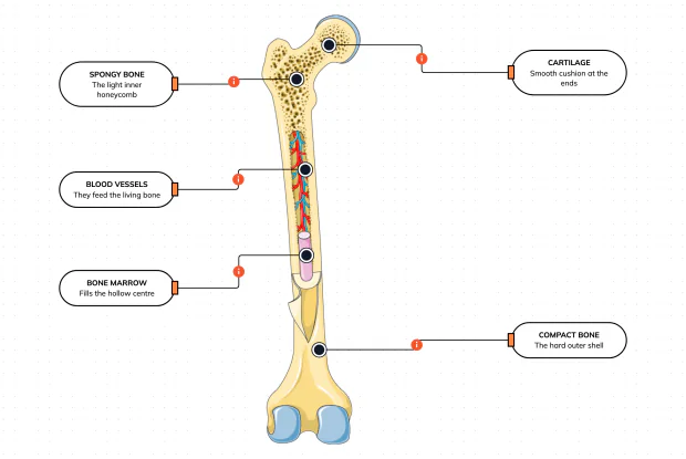
What Is a Bone Made Of?
Cut a bone open and it is nowhere near solid: a hard outer shell, a light honeycomb inside, a hollow centre full of marrow, blood vessels threading through, and a smooth cushion at each end. Learn what each part does — then label them yourself in an interactive playground.
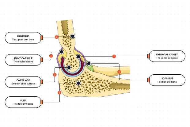
What Is Inside Your Elbow Joint?
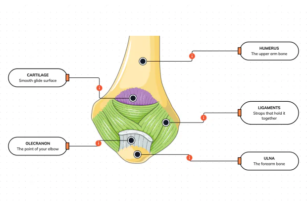
A joint is where two bones meet and move — and it is far more than just the bones. Cut open the elbow and you find smooth cartilage caps, a sealed capsule of slippery fluid, and tough ligaments holding it all together. Learn what each part does, then label them yourself in an interactive playground.
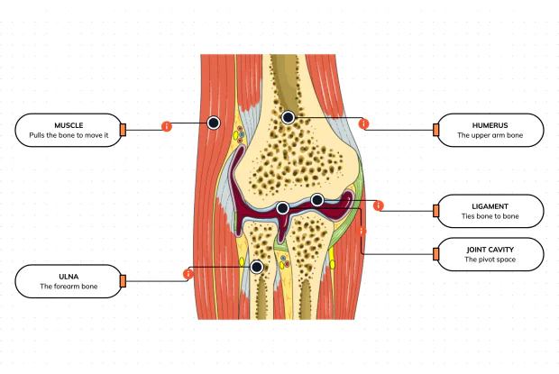
What Makes Your Elbow Bend?
Bones and joints are just the hardware; nothing moves until a muscle pulls. Label the elbow to see the parts that turn a joint into movement — the muscle that pulls, the ligament that holds bone to bone, the two bones it links, and the space they pivot in — then build it yourself in an interactive playground.
What Holds Your Elbow Together?
Your elbow is pulled on all day — by gravity, by every bag you carry, by every time you hang or push. So what stops the two bones from simply coming apart? Look at the back of the elbow and label the ligaments, the bony point, the cartilage and the two bones — and learn what holds the whole joint together.
What Are the Main Bones of the Skull?
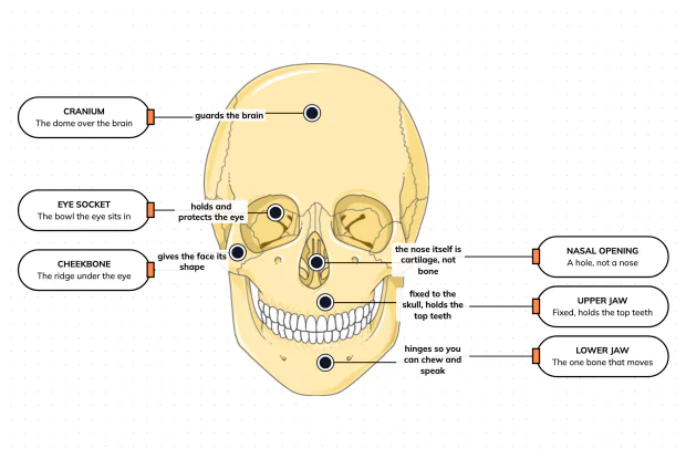
The skull looks like a single bone, but it is really about twenty-two locked together — with just one left free to move. Learn what each main part does, then label a skull yourself in an interactive playground.
What Are the Parts of Your Ear?
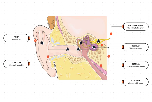
Hearing is a chain of parts passing a sound along, each handing it to the next. Label the pinna, ear canal, eardrum, ossicles, cochlea and auditory nerve, and follow a sound all the way from the air outside to a signal your brain can read — in an interactive playground.
How Does Your Ear Keep You Balanced?
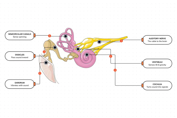
Your ear does a second, hidden job: balance. Inside it sit fluid-filled loops that sense spinning and a chamber that senses tilt and gravity. Label the semicircular canals, vestibule, cochlea, ossicles, eardrum and nerve, and learn how one organ both hears and keeps you upright — in an interactive playground.
What Are the Parts of the Eye You Can See?
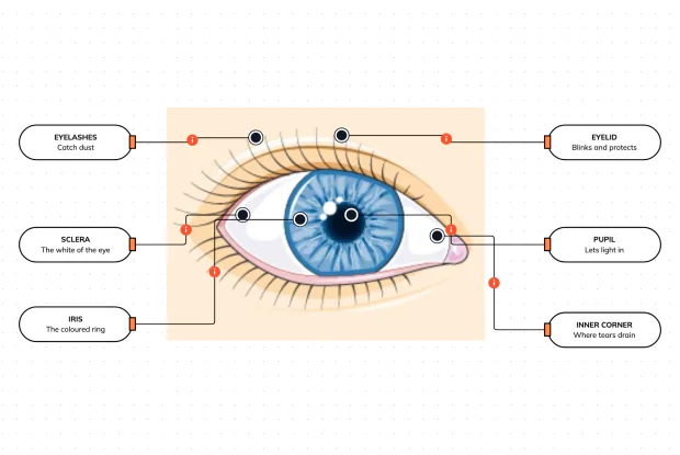
Before you look inside the eye, learn the parts you can already see in a mirror. Label the pupil, iris, sclera, eyelid, eyelashes and the tear-duct corner, and learn what each visible part does to let in light and protect the eye — in an interactive playground.
Where Do Your Tears Come From and Go?
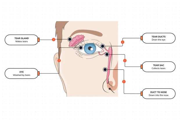
Your eyes make and drain tears all day to stay clean and wet. Label the tear gland, the eye, the tear ducts, the tear sac and the duct to the nose, and follow a tear from where it is made to where it drains — in an interactive playground.
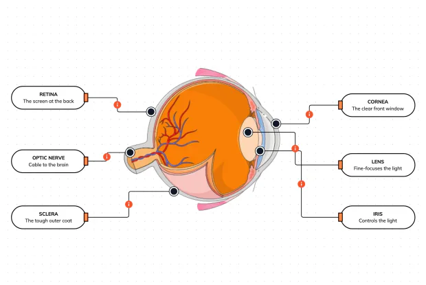
How Does Your Eye Turn Light Into a Picture?
Your eye works like a living camera. Cut it open and label the cornea, lens, iris, retina, optic nerve and sclera, and follow light as it is focused onto a screen at the back of the eye and sent to the brain — in an interactive playground.