How Does Your Eye Bring the World Into Focus?
Root Concept
Light rays from whatever you look at fan out and enter the eye, where the cornea and lens bend them so they meet again at a single focus point. In normal vision that point lands exactly on the retina, forming a sharp image; the lens changes shape to keep near and far objects focused there.
CodePLU Goal
Upgrading Human Mental Models
Learn how to think in Workflows

Concept Development By codeplu.com
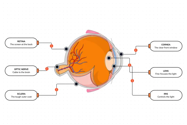
How the eye focuses, each part joined to the job it does — bending light from an object to a single sharp point on the retina
What Does It Actually Mean for Your Eye to Be 'in Focus'?
You say a photo is 'in focus' or 'out of focus' without thinking about what that really means. Here is the idea: from every single point on the thing you are looking at, light sprays outward in all directions, and some of it fans into your eye. To see that point clearly, your eye has to take all those spreading rays and bend them back together so they meet again at one exact point. Do that for every point of the scene at once, and you get a sharp image. Fail to bring the rays back to a point, and everything smears into a blur. That gathering of scattered light back to a point is what focusing is.
Your eye does the bending with two lenses working in series. First the light passes through the cornea, the clear curved window at the front, which bends the rays strongly inward. Then it passes through the lens just behind, which bends them a little more and fine-tunes the aim. Together they take the fan of light rays coming from a point and steer them so they converge — narrowing down to meet at a single focus point deep inside the eye.
The whole trick of good vision is where that focus point lands. In normal vision, the cornea and lens are matched to the length of the eyeball so that the rays meet exactly on the retina, the screen at the back — and a sharp image forms right where the light-sensing cells are waiting. In the playground above you will label the light rays coming in, the cornea and lens that bend them, the focus point where they meet, and the retina they land on — and the label on each connection tells you the job that part does.
How Does the Eye Focus Light Into a Sharp Image?
What does 'in focus' really mean?
It means the light from each point of what you are looking at is brought back together to a single point on the retina. This is the part people rarely picture: light does not travel from an object to your eye in a neat straight beam — it sprays out from every point in all directions, and a small cone of it happens to enter your pupil, spreading as it comes. If nothing bent it, that spreading light would land on the retina as a blurry smear. Focusing is the act of catching that fan of rays and bending it back down to a point. When every point of the scene is re-focused to its own point on the retina, the image is sharp — that is 'in focus'.
What does the cornea do to the light?
The cornea is the clear, strongly-curved dome at the very front of the eye, and it does most of the bending. Because it is curved and sits where the light first arrives — passing from air into the eye — it bends the incoming rays sharply inward, starting them on their way toward a focus point at the back. It is easy to assume the lens does all the focusing, but in fact the fixed cornea provides the bulk of the eye's focusing power; the lens only adds the finishing touch. The cornea's power cannot change, so it always does the same steady, heavy bending, and leaves the adjustable fine-tuning to the lens behind it.
What does the lens add — and how do you focus on near and far things?
The lens sits just behind the cornea and finishes the job the cornea started, bending the rays the last little bit so they meet precisely on the retina. Its special power is that it can change shape: tiny muscles around it squeeze it fatter to focus on something close, or let it thin out to focus on something far away. This constant, automatic reshaping is called accommodation, and it is why you can glance from the words on this page to a view out of the window and have both snap into focus — the lens is adjusting between them in a fraction of a second. The cornea does the big fixed bending; the lens does the flexible, moment-to-moment adjustment that keeps whatever you look at sharp.
Why must the focus point land exactly on the retina?
Because the retina is the only place that can actually record the image — it is the screen covered in light-sensing cells. The cornea and lens always bring the light to a focus somewhere, but a sharp picture only forms where that focus point falls precisely on the retina. In normal vision the eye is perfectly matched: the focusing power of the cornea and lens fits the length of the eyeball so the rays meet right on the retina, and you see clearly. If the eyeball is a touch too long or too short, the light focuses just in front of or behind the retina, and by the time it reaches the screen it has spread out again into a blur — which is short-sightedness or long-sightedness, corrected by adding a lens (glasses) that shifts the focus back onto the retina.
Why is the image on the retina upside-down?
A curious side-effect of focusing light this way is that the image lands on the retina upside-down and back-to-front. It happens because the rays cross over as they pass through the narrow focus: light from the top of the scene ends up at the bottom of the retina, and light from the left ends up on the right. Every lens does this, including a camera's. So the picture painted on the back of your eye is genuinely inverted — yet you see the world the right way up, because your brain has always received it that way and simply interprets it correctly. Your brain, not your eye, decides which way is up; the eye just faithfully focuses the (upside-down) image and sends it on.
Real World Example
Why Can You Focus on Your Finger, Then the Stars, in an Instant?
Focusing feels automatic, but a few everyday moments reveal the machinery — the lens adjusting, the image inverting, and the cornea-plus-lens acting as one camera:
Focusing from your finger to the horizon
The lens changing shape
Hold your finger up close and look at it: it is sharp, and the background is blurry. Now look past it to something far away: the distance snaps into focus and your finger goes blurry. You just watched your lens change shape twice. To focus on the near finger, the muscles around the lens squeezed it fatter to bend the light more strongly; to focus on the distance, they let it thin out. This is accommodation, and it happens in a fraction of a second, all day, without you feeling a thing. It also explains why focusing on close work for hours can tire your eyes — those little muscles are holding the lens squeezed fat the whole time — and why, as the lens stiffens with age, close focusing is the first thing to fail.
The world is upside-down on your retina
The focus point and retina
Because the light rays cross as they squeeze through the focus, the image that actually lands on your retina is upside-down and flipped left-to-right — right now, the picture of this page on the back of your eyes is inverted. You never notice, because your brain has received the world this way since birth and simply reads it correctly; 'up' is whatever your brain has learned to call up. In famous experiments, people who wore glasses that flipped their view upside-down found that after a few days their brains adjusted and the world looked normal again — then flipped once more when they took the glasses off. It is proof that seeing happens in the brain: the eye just focuses the image, inverted and all, and hands it over.
The same trick as a camera
The cornea and lens together
Your eye and a camera solve the exact same problem in the exact same way. Both take the light fanning in from a scene and use curved lenses to bend it back to a focus on a screen — the retina in your eye, the sensor in a camera. Both control the brightness with an adjustable opening (your iris, the camera's aperture). And both must land the focus precisely on the screen, or the picture blurs — which is why a camera has autofocus and your eye has its shape-changing lens. When the eye's focus lands slightly off the retina, the fix is the same one you would use on any camera: add a corrective lens in front. Understanding focusing in the eye is really understanding optics itself — the same rules govern glasses, cameras, telescopes and microscopes.
FAQs
Final Words
Focusing is the heart of seeing, and it is a simple idea: light sprays out from everything you look at, and your eye must bend those scattered rays back together to a point. The cornea does most of that bending as the light enters, the lens fine-tunes it and changes shape to handle near and far, and the rays converge to a focus point. In normal vision that point lands exactly on the retina, so a sharp image forms on the screen and is sent to the brain.
Seeing this explains a surprising amount: why your lens tires after close work, why the image on your retina is upside-down while the world looks upright, and why glasses — a simple extra lens — can fix vision when the focus lands a little off the retina. Your eye is a camera, run by the same optics as every lens ever made. With the outside, the inside, the retina, focusing, and the muscles that aim it, you now understand the eye as a complete instrument.
Continue This Track
This concept is part 12 of The Human Body, Head to Toe.
What Is a Bone Made Of?
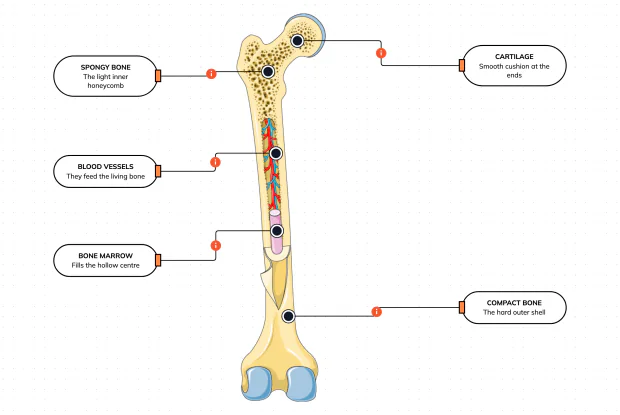
Cut a bone open and it is nowhere near solid: a hard outer shell, a light honeycomb inside, a hollow centre full of marrow, blood vessels threading through, and a smooth cushion at each end. Learn what each part does — then label them yourself in an interactive playground.
What Is Inside Your Elbow Joint?
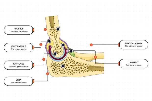
A joint is where two bones meet and move — and it is far more than just the bones. Cut open the elbow and you find smooth cartilage caps, a sealed capsule of slippery fluid, and tough ligaments holding it all together. Learn what each part does, then label them yourself in an interactive playground.
What Makes Your Elbow Bend?
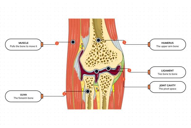
Bones and joints are just the hardware; nothing moves until a muscle pulls. Label the elbow to see the parts that turn a joint into movement — the muscle that pulls, the ligament that holds bone to bone, the two bones it links, and the space they pivot in — then build it yourself in an interactive playground.
What Holds Your Elbow Together?
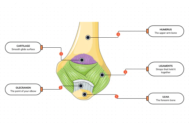
Your elbow is pulled on all day — by gravity, by every bag you carry, by every time you hang or push. So what stops the two bones from simply coming apart? Look at the back of the elbow and label the ligaments, the bony point, the cartilage and the two bones — and learn what holds the whole joint together.
What Are the Main Bones of the Skull?
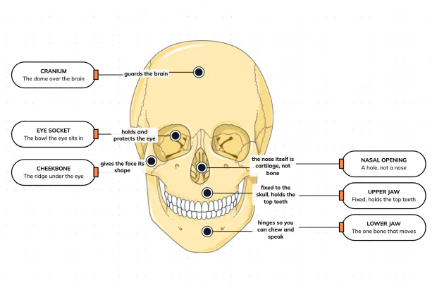
The skull looks like a single bone, but it is really about twenty-two locked together — with just one left free to move. Learn what each main part does, then label a skull yourself in an interactive playground.
What Are the Parts of Your Ear?
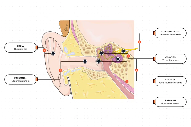
Hearing is a chain of parts passing a sound along, each handing it to the next. Label the pinna, ear canal, eardrum, ossicles, cochlea and auditory nerve, and follow a sound all the way from the air outside to a signal your brain can read — in an interactive playground.
How Does Your Ear Keep You Balanced?
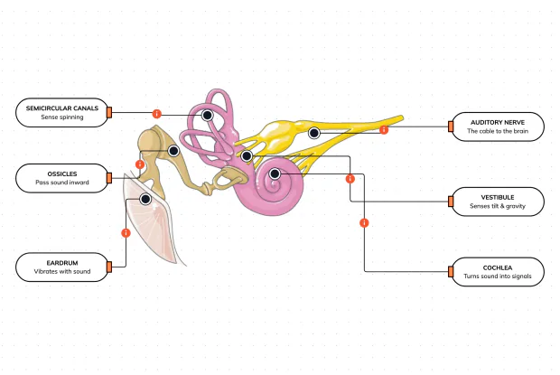
Your ear does a second, hidden job: balance. Inside it sit fluid-filled loops that sense spinning and a chamber that senses tilt and gravity. Label the semicircular canals, vestibule, cochlea, ossicles, eardrum and nerve, and learn how one organ both hears and keeps you upright — in an interactive playground.
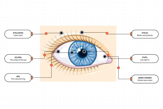
What Are the Parts of the Eye You Can See?
Before you look inside the eye, learn the parts you can already see in a mirror. Label the pupil, iris, sclera, eyelid, eyelashes and the tear-duct corner, and learn what each visible part does to let in light and protect the eye — in an interactive playground.
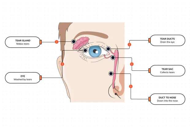
Where Do Your Tears Come From and Go?
Your eyes make and drain tears all day to stay clean and wet. Label the tear gland, the eye, the tear ducts, the tear sac and the duct to the nose, and follow a tear from where it is made to where it drains — in an interactive playground.
How Does Your Eye Turn Light Into a Picture?
Your eye works like a living camera. Cut it open and label the cornea, lens, iris, retina, optic nerve and sclera, and follow light as it is focused onto a screen at the back of the eye and sent to the brain — in an interactive playground.