What Are the Lobes of the Lungs?
Root Concept
Each lung is divided by deep grooves called fissures into sections called lobes. The right lung has three lobes — upper, middle and lower — while the left lung has only two — upper and lower — because space on the left is taken up by the heart.
CodePLU Goal
Upgrading Human Mental Models
Learn how to think in Workflows

Concept Development By codeplu.com
All five lung lobes labelled — the right lung's upper, middle and lower, and the left lung's upper and lower, divided by their fissures
Why Isn't Each Lung Just One Solid Piece?
From the outside a lung looks like one smooth, spongy bag, but it is not a single piece. Each lung is divided by deep grooves, called fissures, into separate sections called lobes. It is a bit like an orange split into segments — still one fruit, but made of clear parts. These lobes are the main building blocks of each lung.
Here is the surprising bit: the two lungs are not the same. The right lung is split into three lobes — an upper, a middle and a lower — by two fissures. The left lung has only two lobes — an upper and a lower — divided by a single fissure. So altogether you have five lung lobes: three on the right, two on the left.
The reason the left lung has one fewer lobe is that it has to share its side of the chest with the heart, which sits slightly to the left. The left lung even has a scooped-out notch to make room for it. In the playground you will label all five lobes — the right lung's three and the left lung's two — using the fissures to tell them apart.
How Are the Lungs Divided Into Lobes?
What is a lobe, and what is a fissure?
A lobe is a large, distinct section of a lung, and the deep groove that separates one lobe from another is called a fissure. If you ran your finger along the surface of a lung, you would feel these fissures as clefts dividing it into pieces, rather like the segments of an orange. The lobes are not separate organs — the whole lung still works together as one — but each lobe is a fairly independent unit, with its own branch of the airway (a bronchus) delivering air to it and its own blood supply. Dividing the lung into lobes this way has a real advantage: if disease strikes, it often stays within one lobe instead of spreading through the whole lung, and a surgeon can sometimes remove just one affected lobe and leave the rest working.
The right lung: three lobes
The right lung is the larger of the two, and it is divided into three lobes: the upper (or superior) lobe at the top, the middle lobe in the front-lower part, and the lower (or inferior) lobe making up most of the bottom and back. Two fissures do the dividing. The oblique fissure runs diagonally and separates the lower lobe from the two above it, and the horizontal fissure runs across the front to split the upper lobe from the middle lobe. The middle lobe is the smallest of the three and is special: it is the one lobe that only the right lung has. So whenever you need to remember which lung has the extra lobe, it is the right — three lobes on the right, thanks to that second, horizontal fissure.
The left lung: two lobes and room for the heart
The left lung has just two lobes — an upper (superior) lobe and a lower (inferior) lobe — divided by a single oblique fissure running diagonally down it. It has no middle lobe and no horizontal fissure. The reason is simple: the heart sits slightly to the left of the centre of the chest, so the left lung has less room and is a little smaller than the right. The left lung even carries a curved dent on its inner-front edge, called the cardiac notch, which cups around the heart to make space for it. So the left lung's two-lobe design is really a compromise with its neighbour: it gives up a lobe, and part of its shape, so the heart has somewhere to sit right beside it.
Real World Example
Why Knowing the Lobes Matters
The lobes are not just neat anatomy; they shape how lung problems are found and treated. Here are three:
Pneumonia in one lobe
Infection often stays inside a single lobe
When someone gets pneumonia, an infection of the lung, it frequently settles in just one lobe rather than filling the whole lung — doctors might describe it as 'right-lower-lobe pneumonia', for example. This happens partly because each lobe is a fairly self-contained unit with its own airway branch, so an infection travelling down one bronchus tends to flood that lobe and stop at its fissures. Knowing exactly which lobe is affected helps doctors listen to the right spot with a stethoscope, point to it on a chest X-ray, and track whether it is clearing up. So the lobes give illness an address: naming the lobe tells everyone precisely where the problem is.
Removing just one lobe
A lobectomy takes a lobe and spares the rest
Because each lobe is a semi-independent unit, surgeons can sometimes remove a single diseased lobe — an operation called a lobectomy — while leaving the other lobes to carry on breathing. If a tumour is contained within, say, the right upper lobe, taking out that one lobe may treat it without sacrificing the whole lung. The patient breathes on the remaining lobes, which take over the work. This is only possible because the lung is built in lobes with their own airways and blood supply, meeting at the fissures. It is a clear, life-saving example of why the body's habit of building organs from separable parts is so useful.
Which lung has room for the heart
The left's two lobes explain the chest's layout
If you are ever asked why the two lungs are different, the answer is the heart. The heart sits just left of centre, so the left lung is smaller, carries a cardiac notch, and makes do with two lobes, while the roomier right lung has three. This asymmetry is easy to see on any chest scan and explains lots of everyday anatomy: why the right lung is a little bigger, why the windpipe's right branch is more upright (so inhaled objects more often end up in the right lung), and why doctors always keep track of left versus right. The number of lobes — three on the right, two on the left — is the simplest sign of how the lungs share the chest with the heart.
FAQs
Final Words
Each lung is split by fissures into lobes: the right lung has three — upper, middle and lower — divided by an oblique and a horizontal fissure, while the left lung has just two — upper and lower — divided by a single oblique fissure. That makes five lobes in all, and the middle lobe is the one feature unique to the right lung. The left lung has fewer because it shares its side of the chest with the heart, even carving out a cardiac notch to fit it.
These lobes are more than tidy anatomy: each is a semi-independent unit with its own airway and blood supply, which is why infections often stay in one lobe and why a single lobe can be removed in surgery while the rest keep breathing. Three on the right, two on the left — a simple count that captures how the lungs are built and how they share the chest with the heart.
Continue This Track
This concept is part 56 of The Human Body, Head to Toe.
What Is a Bone Made Of?
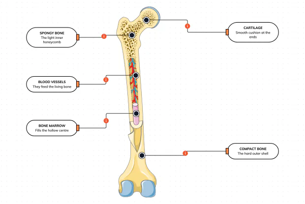
Cut a bone open and it is nowhere near solid: a hard outer shell, a light honeycomb inside, a hollow centre full of marrow, blood vessels threading through, and a smooth cushion at each end. Learn what each part does — then label them yourself in an interactive playground.
What Is Inside Your Elbow Joint?
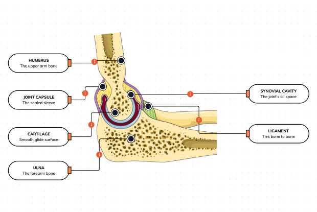
A joint is where two bones meet and move — and it is far more than just the bones. Cut open the elbow and you find smooth cartilage caps, a sealed capsule of slippery fluid, and tough ligaments holding it all together. Learn what each part does, then label them yourself in an interactive playground.
What Makes Your Elbow Bend?
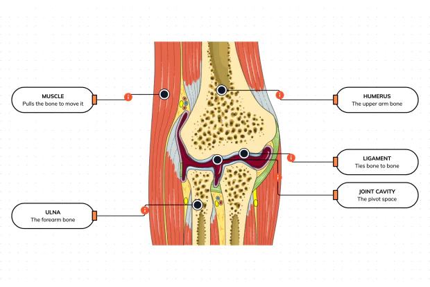
Bones and joints are just the hardware; nothing moves until a muscle pulls. Label the elbow to see the parts that turn a joint into movement — the muscle that pulls, the ligament that holds bone to bone, the two bones it links, and the space they pivot in — then build it yourself in an interactive playground.
What Holds Your Elbow Together?
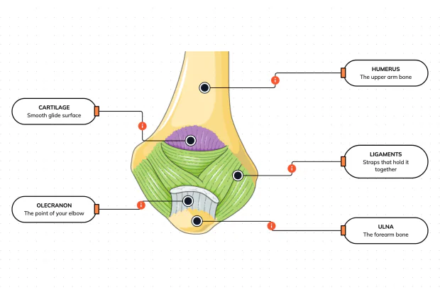
Your elbow is pulled on all day — by gravity, by every bag you carry, by every time you hang or push. So what stops the two bones from simply coming apart? Look at the back of the elbow and label the ligaments, the bony point, the cartilage and the two bones — and learn what holds the whole joint together.
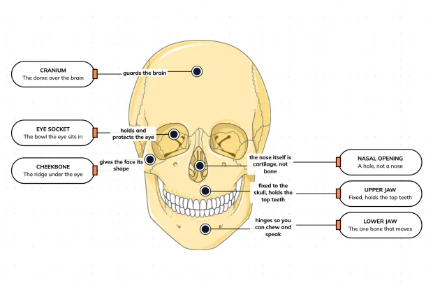
What Are the Main Bones of the Skull?
The skull looks like a single bone, but it is really about twenty-two locked together — with just one left free to move. Learn what each main part does, then label a skull yourself in an interactive playground.
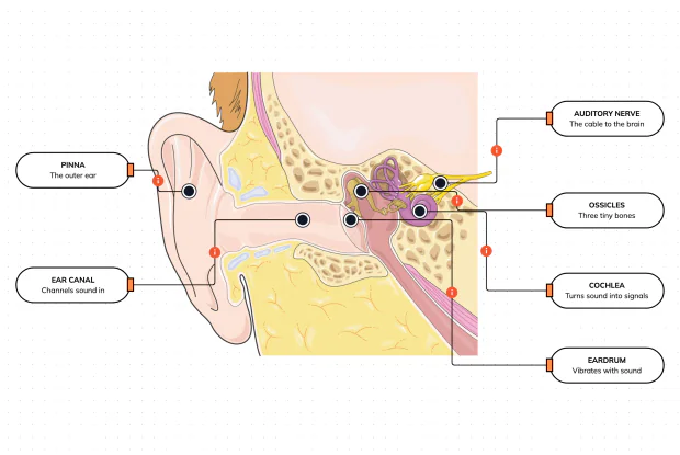
What Are the Parts of Your Ear?
Hearing is a chain of parts passing a sound along, each handing it to the next. Label the pinna, ear canal, eardrum, ossicles, cochlea and auditory nerve, and follow a sound all the way from the air outside to a signal your brain can read — in an interactive playground.
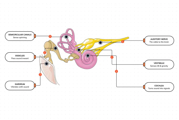
How Does Your Ear Keep You Balanced?
Your ear does a second, hidden job: balance. Inside it sit fluid-filled loops that sense spinning and a chamber that senses tilt and gravity. Label the semicircular canals, vestibule, cochlea, ossicles, eardrum and nerve, and learn how one organ both hears and keeps you upright — in an interactive playground.
What Are the Parts of the Eye You Can See?
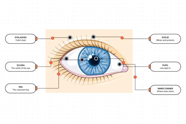
Before you look inside the eye, learn the parts you can already see in a mirror. Label the pupil, iris, sclera, eyelid, eyelashes and the tear-duct corner, and learn what each visible part does to let in light and protect the eye — in an interactive playground.
Where Do Your Tears Come From and Go?
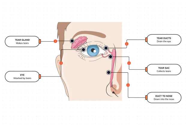
Your eyes make and drain tears all day to stay clean and wet. Label the tear gland, the eye, the tear ducts, the tear sac and the duct to the nose, and follow a tear from where it is made to where it drains — in an interactive playground.
How Does Your Eye Turn Light Into a Picture?
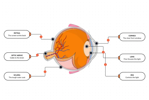
Your eye works like a living camera. Cut it open and label the cornea, lens, iris, retina, optic nerve and sclera, and follow light as it is focused onto a screen at the back of the eye and sent to the brain — in an interactive playground.