How Does the Liver Work, Up Close?
Root Concept
The liver is made of thousands of tiny units called lobules. In each, blood enters from a portal vein and hepatic artery at the edges, trickles through leaky channels called sinusoids past rows of liver cells (hepatocytes) that process and clean it, and the cleaned blood drains out through a central vein. Bile made by the cells flows the other way into a bile duct.
CodePLU Goal
Upgrading Human Mental Models
Learn how to think in Workflows

Concept Development By codeplu.com
Inside a liver lobule, each part joined to the job it does as blood flows past the worker cells to be cleaned and bile is drawn off
How Can One Organ Process Everything in Your Blood?
The whole-organ view of the liver tells you what it does — process nutrients, clean the blood, make bile — but not how one lump of tissue manages such an enormous chemical workload. The secret is in its structure: the liver is not a solid block but is built from thousands of tiny, identical units called lobules, each a miniature processing station. Zoom in on one, and the liver's genius becomes clear — it is designed so that every drop of blood is forced to trickle slowly past its worker cells, giving them maximum chance to work on it.
A lobule is arranged around a drain in the middle: the central vein at the centre. Radiating out from it are neat rows of liver cells, the hepatocytes — these are the actual workers that do the liver's chemistry. Running between the rows of cells are the sinusoids: special leaky blood channels, so open and porous that the blood almost bathes the cells directly rather than being sealed inside tight vessels. This is the whole trick — the blood is put into intimate, slow contact with the cells that must process it.
Blood enters at the edges of the lobule, where three vessels run together (the 'portal triad'): the portal vein brings nutrient-rich blood from the gut, and the hepatic artery brings oxygen-rich blood. Both trickle inward through the sinusoids, past the rows of cells, to be collected and drained away by the central vein. Meanwhile the cells make bile, which flows the opposite way, out to a small bile duct. In the playground above you will label the liver cells, sinusoid, central vein, portal vein, hepatic artery and bile duct — and the label on each connection tells you the job that part does.
What Does Each Part of the Liver Lobule Do?
What is a liver lobule?
A lobule is one tiny building-block of the liver — the whole organ is made of thousands of them packed together, each doing the same job on its own little share of the blood. Think of the liver less like a single factory and more like a warehouse full of thousands of identical small workstations. Each lobule is roughly a cylinder built around a central drain vein, with liver cells stacked in rows around it and blood channels running between them. This repeating design is powerful for two reasons: it spreads the blood out so that every drop passes close to worker cells, and it means the liver can lose or damage many lobules and keep working, because the rest simply carry on. Understanding one lobule is understanding how the entire liver works.
What do the liver cells (hepatocytes) do?
The liver cells — hepatocytes — are the workers that actually perform the liver's hundreds of chemical jobs. As blood flows slowly past them, they reach into it and get to work: pulling out nutrients to store or convert, grabbing toxins, alcohol and drug by-products to break down and neutralise, building proteins to release back into the blood, and manufacturing bile. Every famous thing the liver 'does' is really these cells doing it, multiplied by billions. They are arranged in thin rows exactly one or two cells thick, so that no cell is ever far from the blood it works on — a layout that gives the maximum number of cells direct access to the passing blood. They are also the cells that let the liver regrow, dividing to replace tissue that is lost.
What is a sinusoid?
A sinusoid is the special blood channel that runs between the rows of liver cells — and it is unlike an ordinary blood vessel. Normal vessels have sealed walls that keep blood neatly inside; sinusoids are deliberately leaky, with gaps in their walls, so that the blood plasma can seep out and bathe the liver cells almost directly. This is essential: for the cells to process substances in the blood, they need to actually touch it, not be walled off from it. So the sinusoid is where the exchange happens — nutrients, toxins and oxygen pass from the blood into the cells, and proteins and processed substances pass back out. The blood also flows through them slowly, which gives the cells time to do their work as it drifts past.
What does the central vein do?
The central vein sits at the very middle of the lobule and is the drain: it collects the blood after it has passed through all the sinusoids and been worked on by the cells, and carries it away out of the liver. The flow is inward-then-out: blood enters at the edges, trickles through the sinusoids toward the centre, and by the time it reaches the central vein it has been cleaned, its nutrients processed, its toxins removed. All the central veins from all the lobules join up into larger veins that eventually return the processed blood to the heart, to be sent on to the rest of the body. So the central vein marks the end of the blood's journey through the lobule — the exit for freshly-serviced blood.
Where does the blood come in?
At the outer edges of the lobule, where two supply vessels run together. The portal vein branch brings nutrient-rich blood straight from the gut — full of everything just absorbed from a meal — which is exactly what the liver cells need to process. The hepatic artery branch brings oxygen-rich blood from the heart to keep the hardworking cells alive. The two streams mix and trickle inward together through the sinusoids, so each cell gets both the raw materials to work on (from the portal vein) and the oxygen to power the work (from the artery). This pairing of the two supplies at the lobule's edge, along with a bile duct, is such a consistent feature that the trio is known as the 'portal triad'.
Where does the bile go?
The bile the liver cells make cannot mix into the blood, so it needs its own separate drainage — and it flows in the opposite direction to the blood. While blood trickles inward toward the central vein, the bile is collected into tiny channels running outward between the cells, toward the edge of the lobule, where it empties into a small bile duct. From there, bile ducts from all the lobules join into bigger and bigger ducts, eventually forming the main bile duct that carries bile to the gallbladder and intestine. So a lobule actually has two-way traffic: blood moving in one direction to be cleaned, and bile moving the other way to be shipped out — kept carefully separate the whole time.
Real World Example
Why Is the Liver So Good at Its Job?
The clever structure of the lobule explains how the liver achieves so much, and why it is so resilient. Three features of the design tell the story:
Blood flows slowly past every cell
The sinusoids
The liver's biggest challenge is contact: to process the blood, its cells must physically touch it, and there is a lot of blood to get through. The sinusoids solve this beautifully. Instead of sealed pipes rushing blood past, they are wide, leaky channels that let the blood ooze out and bathe the cells directly, and they slow the flow right down so the blood dawdles past rank after rank of hepatocytes. With the cells stacked only one or two deep along every channel, virtually no drop of blood escapes without being worked on. It is the same principle as the tiny folds of the small intestine or the branching of the lungs: maximise the contact area and slow things down, so a huge amount of processing happens in a small space.
Two-way traffic, kept apart
Blood and bile flowing opposite ways
A liver cell has to do two shipping jobs at once: take things out of the blood, and send bile away — and bile must never leak into the blood. The lobule handles this with elegant counter-flow. Blood travels inward, from the edges toward the central vein, getting cleaner as it goes; bile travels outward, from the cells toward the bile duct at the edge, in tiny separate channels. The two never mix, running in opposite directions through the same tissue like inbound and outbound lanes of a motorway. This is why a blockage in the bile drainage is so serious: with the bile's exit route blocked, it can back up into the blood, which is exactly what turns the skin yellow in jaundice.
Thousands of identical units
The repeating lobule design
Because the liver is built from thousands of identical lobules rather than as one big structure, it is remarkably tough. Damage or destroy a batch of lobules — through injury, infection or toxins — and the rest simply keep working, so the liver can lose a surprising amount of tissue and still do its job. Combined with the hepatocytes' rare ability to divide and regrow, this repeating design is why a healthy liver can regenerate after part of it is removed, and why a person can donate a piece of their liver and have it grow back. It is the same logic as a data centre built from many identical servers: no single point of failure, and easy to rebuild unit by unit. The lobule is the liver's secret to being both powerful and resilient.
FAQs
Final Words
The liver's power comes from its structure. It is built from thousands of tiny repeating lobules, and inside each one the design forces every drop of blood to trickle slowly through leaky sinusoids, past rows of worker cells (hepatocytes) that clean it and make bile. Blood enters at the edges from the portal vein and hepatic artery, is processed as it flows inward, and drains out cleaned through the central vein — while bile flows the opposite way to a bile duct, never mixing.
This clever layout — huge contact area, slow flow, two-way traffic and endless identical units — is how one organ processes everything you eat, drink and absorb, and why it can take damage and regrow. You have now seen the liver both as a whole organ and down at the scale where its work actually happens. It is a perfect example of a theme running through the whole body: zoom in, and big jobs turn out to be done by many tiny, well-designed repeating units.
Continue This Track
This concept is part 31 of The Human Body, Head to Toe.
What Is a Bone Made Of?
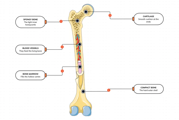
Cut a bone open and it is nowhere near solid: a hard outer shell, a light honeycomb inside, a hollow centre full of marrow, blood vessels threading through, and a smooth cushion at each end. Learn what each part does — then label them yourself in an interactive playground.
What Is Inside Your Elbow Joint?
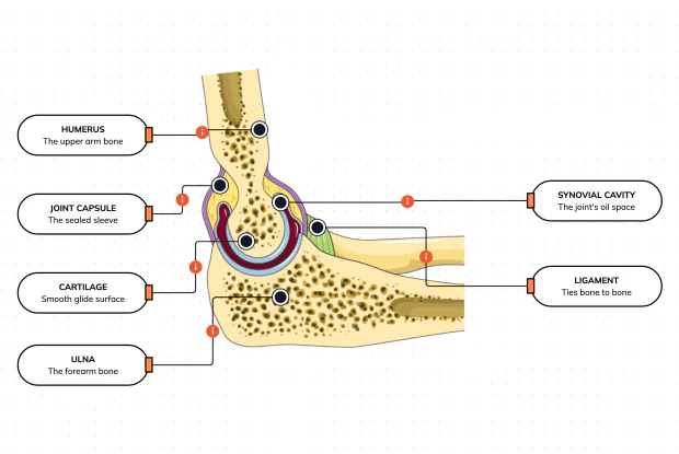
A joint is where two bones meet and move — and it is far more than just the bones. Cut open the elbow and you find smooth cartilage caps, a sealed capsule of slippery fluid, and tough ligaments holding it all together. Learn what each part does, then label them yourself in an interactive playground.
What Makes Your Elbow Bend?
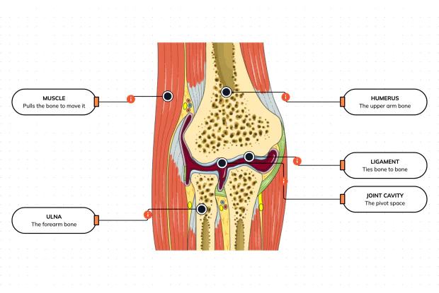
Bones and joints are just the hardware; nothing moves until a muscle pulls. Label the elbow to see the parts that turn a joint into movement — the muscle that pulls, the ligament that holds bone to bone, the two bones it links, and the space they pivot in — then build it yourself in an interactive playground.
What Holds Your Elbow Together?
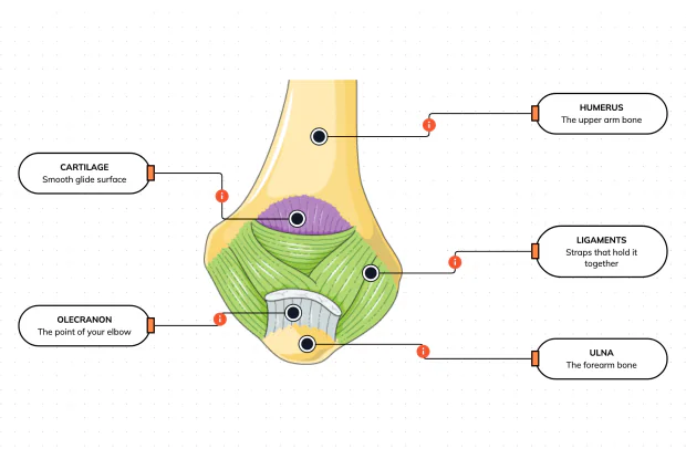
Your elbow is pulled on all day — by gravity, by every bag you carry, by every time you hang or push. So what stops the two bones from simply coming apart? Look at the back of the elbow and label the ligaments, the bony point, the cartilage and the two bones — and learn what holds the whole joint together.
What Are the Main Bones of the Skull?
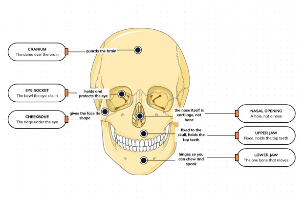
The skull looks like a single bone, but it is really about twenty-two locked together — with just one left free to move. Learn what each main part does, then label a skull yourself in an interactive playground.
What Are the Parts of Your Ear?
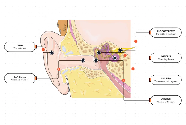
Hearing is a chain of parts passing a sound along, each handing it to the next. Label the pinna, ear canal, eardrum, ossicles, cochlea and auditory nerve, and follow a sound all the way from the air outside to a signal your brain can read — in an interactive playground.
How Does Your Ear Keep You Balanced?
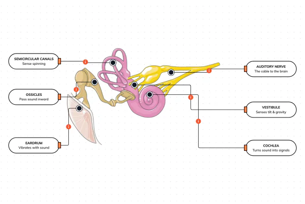
Your ear does a second, hidden job: balance. Inside it sit fluid-filled loops that sense spinning and a chamber that senses tilt and gravity. Label the semicircular canals, vestibule, cochlea, ossicles, eardrum and nerve, and learn how one organ both hears and keeps you upright — in an interactive playground.
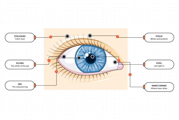
What Are the Parts of the Eye You Can See?
Before you look inside the eye, learn the parts you can already see in a mirror. Label the pupil, iris, sclera, eyelid, eyelashes and the tear-duct corner, and learn what each visible part does to let in light and protect the eye — in an interactive playground.
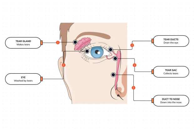
Where Do Your Tears Come From and Go?
Your eyes make and drain tears all day to stay clean and wet. Label the tear gland, the eye, the tear ducts, the tear sac and the duct to the nose, and follow a tear from where it is made to where it drains — in an interactive playground.
How Does Your Eye Turn Light Into a Picture?
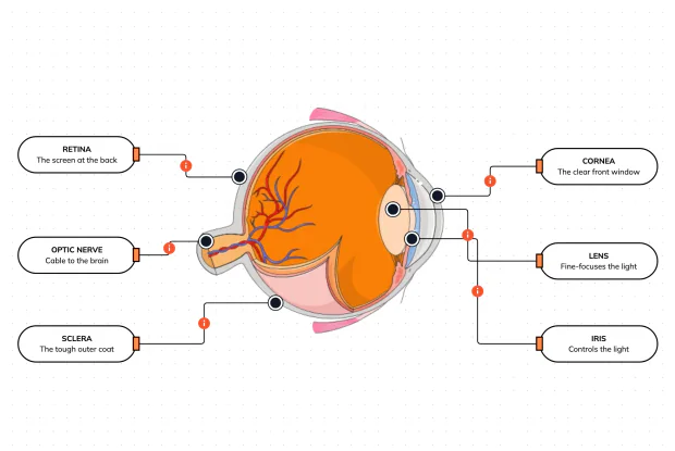
Your eye works like a living camera. Cut it open and label the cornea, lens, iris, retina, optic nerve and sclera, and follow light as it is focused onto a screen at the back of the eye and sent to the brain — in an interactive playground.