How Does Your Gut Absorb Food Into Your Blood?
Root Concept
The small intestine's lining is covered in tiny finger-like villi that hugely increase the absorbing surface. Each villus is coated in absorptive cells whose surface carries even tinier microvilli; digested nutrients cross these cells into a central lacteal (which absorbs fat) and blood capillaries (which absorb sugars and amino acids), while goblet cells make protective mucus.
CodePLU Goal
Upgrading Human Mental Models
Learn how to think in Workflows

Concept Development By codeplu.com
An intestinal villus, each part joined to the job it does — taking nutrients in and shipping them out into the blood and lymph
How Does a Nutrient Get From Your Gut Into Your Blood?
By the time food reaches the middle of the small intestine, it has been broken down into its smallest pieces — sugars, amino acids, fat droplets, vitamins. But those nutrients are still inside the gut, which is really just a tube passing through your body; to actually feed you, they have to cross the gut wall and get into your blood. That final, crucial crossing happens here, at the tiny finger-like bumps that carpet the intestine's lining: the villi. Each is less than a millimetre tall, and there are millions of them, turning the inner surface into something like a deep-pile velvet carpet.
The reason for all these fingers is surface area — the same theme as the whole small intestine, taken to its extreme. Each villus is coated in a layer of tall absorptive cells, and the surface of every one of those cells is itself covered in even tinier hairs called microvilli. So the surface is folded at three scales at once — the whole intestine is folded into ridges, the ridges sprout villi, and the villi's cells sprout microvilli — which is how the gut ends up with the absorbing area of a tennis court packed inside your belly.
Inside each villus is the delivery system. Running up its core is a lacteal — a tiny lymph vessel that soaks up digested fat — surrounded by a network of blood capillaries that soak up sugars and amino acids. Nutrients pass through the absorptive cells and into one or other of these, to be carried off and distributed around the body. Scattered among the absorptive cells are goblet cells that make protective mucus. In the playground above you will label the microvilli, absorptive cells, lacteal, blood capillaries and goblet cell — and the label on each connection tells you the job that part does.
How Does a Villus Absorb Nutrients?
What is a villus, and why are there so many?
A villus is a tiny, finger-like projection of the intestinal lining — less than a millimetre tall — and the small intestine has millions of them, standing side by side like the pile of a plush carpet. Their whole purpose is to increase surface area for absorption. A smooth tube of the same length would have a fairly small inner surface, but covering that surface in millions of tiny fingers multiplies the area enormously, because every finger adds its own sides and tip to the total. This is the second level of the intestine's folding trick: the tube is already thrown into large circular folds, and then those folds are carpeted with villi. More surface means more room for nutrients to cross into the body, so the meal can be absorbed as completely as possible before it moves on.
What are the absorptive cells and microvilli?
The surface of each villus is a single layer of tall, column-shaped absorptive cells, and these are the actual gatekeepers: digested nutrients pass through them to get from the gut into the villus. But the surface trick goes one level deeper still. The top of each absorptive cell — the side facing the gut — is covered in a dense fuzz of microscopic projections called microvilli, together forming what is called the brush border. These multiply the absorbing surface yet again, so that even a single cell has a hugely magnified area to take nutrients in through. It is folding upon folding upon folding: intestine into folds, folds into villi, and villi's cells into microvilli — three nested levels, each multiplying the last, all to maximise the surface where absorption happens.
What does the lacteal do?
Running up the centre of each villus is a lacteal — a tiny vessel that belongs not to the blood system but to the lymphatic system, and its special job is to absorb digested fat. Fat is handled differently from other nutrients: once broken into droplets and taken up by the absorptive cells, it is repackaged and released into the lacteal rather than straight into the blood. The lacteal collects this fatty fluid (which looks milky, giving the lacteal its name, from the Latin for milk) and carries it away through the lymph vessels, which eventually empty it into the bloodstream near the heart. So fat takes a scenic, separate route out of the gut — into the lacteal, through the lymph system, and only then into the blood. The lacteal is the fat's private exit from each villus.
What do the blood capillaries do?
Wrapped around the lacteal, inside each villus, is a fine mesh of blood capillaries — and these absorb the water-soluble nutrients: the sugars from carbohydrates and the amino acids from proteins. As these nutrients pass through the absorptive cells, they cross into this capillary network and are carried away in the blood. And that blood does not go straight to the body — it is gathered up and sent first to the liver (through the portal vein you met earlier), so the liver can process and check everything before it circulates. So each villus has two shipping routes leaving it: the blood capillaries for sugars and amino acids, and the lacteal for fats. Together they carry the whole absorbed meal out of the gut and into the body.
What is the goblet cell for?
Dotted among the tall absorptive cells are rounded cells shaped a little like a wine goblet — the goblet cells — and their job is to make mucus. They release slippery mucus onto the surface of the lining, and this does two important things: it lubricates, helping the food slide smoothly along, and it protects, coating the delicate absorbing cells so they are not damaged by the acids, enzymes and rough contents passing over them. The lining of the whole gut has goblet cells for this reason, and they become even more numerous further along, in the large intestine, where the drying leftovers need extra lubrication. So while the absorptive cells do the taking-in, the goblet cells keep the surface safe and slippery — a small but essential supporting role on the absorbing surface.
Real World Example
Why Does the Same Meal Leave Your Gut by Two Different Doors?
A single villus quietly performs two clever tricks at once — maximising surface, and sorting nutrients. Seeing them explains how absorption works and what happens when the villi are damaged:
Surface upon surface upon surface
Villi and microvilli
The gut's challenge is to absorb a whole meal, and its answer is to pack in as much absorbing surface as physically possible — by folding at three scales at once. The intestine's lining is thrown into large circular folds; those folds are carpeted with millions of tiny villi; and each villus's cells are tipped with countless even-tinier microvilli. Each level multiplies the last, so a modest length of tube ends up with an absorbing surface roughly the size of a tennis court, all hidden inside your belly. It is the same principle the lungs use to pack in surface for absorbing oxygen: when you need a huge area in a small space, you fold, and then fold the folds, and then fold those. The villus is that principle made visible.
Two exits for two kinds of food
The lacteal and the capillaries
Here is the surprising part: your villus sorts nutrients and ships them out by two different routes. Sugars and amino acids — the water-soluble nutrients — pass into the blood capillaries and are carried off in the blood, straight to the liver for processing. Fats, which do not mix with water, are handled separately: they are packaged up and released into the central lacteal, a lymph vessel, and travel through the lymph system before finally joining the blood near the heart. So a single mouthful is split at the villus into two streams leaving by two doors — the fatty part through the lymph, the rest through the blood. It is a neat solution to the problem that fat and water do not mix, built right into the structure of every villus.
When the villi are flattened
Damaged absorbing surface
Because absorption depends entirely on this vast folded surface, damaging the villi is serious. In coeliac disease, for example, the immune system reacts to gluten and attacks the lining of the small intestine, gradually wearing the villi down until they are flattened. With the fingers gone, the absorbing surface collapses from a tennis court to something far smaller — and even though the person is eating normally, they cannot absorb enough nutrients, leading to tiredness, weight loss and deficiencies. It is a striking demonstration of how much the body relies on those tiny fingers: lose the villi, and a healthy diet is no longer enough. Removing the gluten lets the villi grow back and absorption recovers — which is why the treatment is a strict gluten-free diet, not a medicine.
FAQs
Final Words
The villi are where a meal finally becomes part of you. The small intestine's lining is carpeted with millions of these tiny fingers, each coated in absorptive cells whose surface bristles with microvilli — folding upon folding upon folding, building an absorbing surface the size of a tennis court. As nutrients cross the absorptive cells, they are sorted into two exits: fats into the central lacteal and the lymph, sugars and amino acids into the blood capillaries and on to the liver, while goblet cells keep the surface protected with mucus.
This is the whole point of digestion, happening at the smallest scale: everything upstream — chewing, the stomach's acid, the duodenum's juices, the intestine's coils — exists to deliver a meal, broken down, to these tiny fingers where it can cross into your body. It also shows how fragile that surface is: flatten the villi, as coeliac disease does, and a full plate can no longer feed you. Next the tour follows the leftovers onward into the large intestine.
Continue This Track
This concept is part 37 of The Human Body, Head to Toe.
What Is a Bone Made Of?
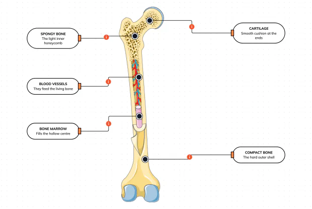
Cut a bone open and it is nowhere near solid: a hard outer shell, a light honeycomb inside, a hollow centre full of marrow, blood vessels threading through, and a smooth cushion at each end. Learn what each part does — then label them yourself in an interactive playground.
What Is Inside Your Elbow Joint?
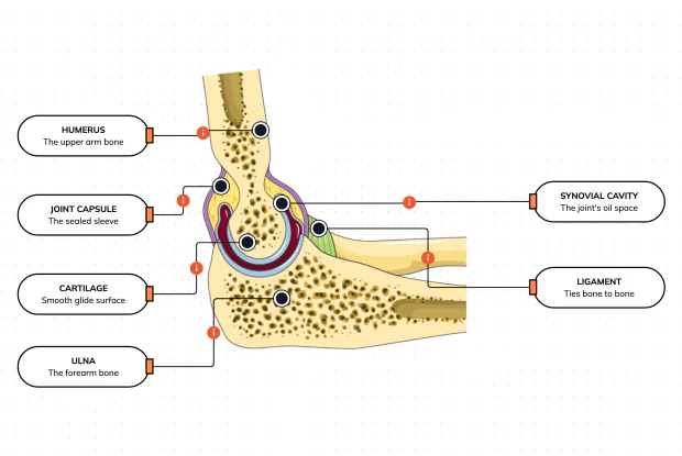
A joint is where two bones meet and move — and it is far more than just the bones. Cut open the elbow and you find smooth cartilage caps, a sealed capsule of slippery fluid, and tough ligaments holding it all together. Learn what each part does, then label them yourself in an interactive playground.
What Makes Your Elbow Bend?
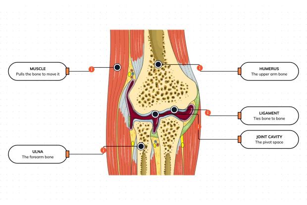
Bones and joints are just the hardware; nothing moves until a muscle pulls. Label the elbow to see the parts that turn a joint into movement — the muscle that pulls, the ligament that holds bone to bone, the two bones it links, and the space they pivot in — then build it yourself in an interactive playground.
What Holds Your Elbow Together?
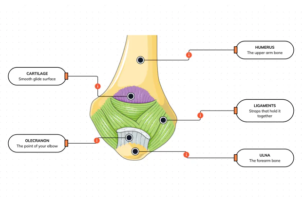
Your elbow is pulled on all day — by gravity, by every bag you carry, by every time you hang or push. So what stops the two bones from simply coming apart? Look at the back of the elbow and label the ligaments, the bony point, the cartilage and the two bones — and learn what holds the whole joint together.
What Are the Main Bones of the Skull?
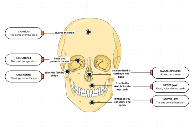
The skull looks like a single bone, but it is really about twenty-two locked together — with just one left free to move. Learn what each main part does, then label a skull yourself in an interactive playground.
What Are the Parts of Your Ear?
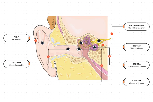
Hearing is a chain of parts passing a sound along, each handing it to the next. Label the pinna, ear canal, eardrum, ossicles, cochlea and auditory nerve, and follow a sound all the way from the air outside to a signal your brain can read — in an interactive playground.
How Does Your Ear Keep You Balanced?
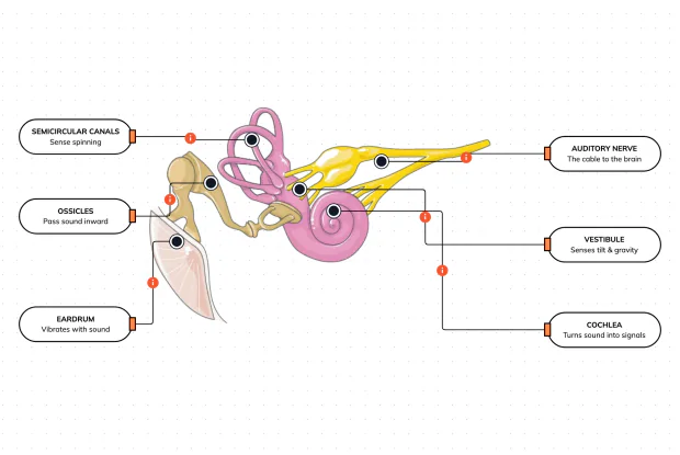
Your ear does a second, hidden job: balance. Inside it sit fluid-filled loops that sense spinning and a chamber that senses tilt and gravity. Label the semicircular canals, vestibule, cochlea, ossicles, eardrum and nerve, and learn how one organ both hears and keeps you upright — in an interactive playground.
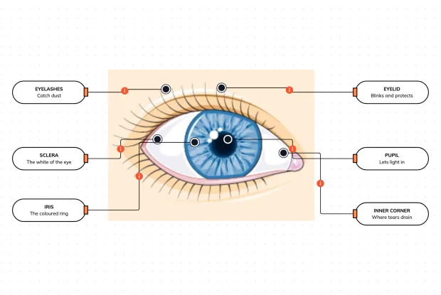
What Are the Parts of the Eye You Can See?
Before you look inside the eye, learn the parts you can already see in a mirror. Label the pupil, iris, sclera, eyelid, eyelashes and the tear-duct corner, and learn what each visible part does to let in light and protect the eye — in an interactive playground.
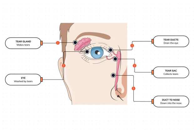
Where Do Your Tears Come From and Go?
Your eyes make and drain tears all day to stay clean and wet. Label the tear gland, the eye, the tear ducts, the tear sac and the duct to the nose, and follow a tear from where it is made to where it drains — in an interactive playground.
How Does Your Eye Turn Light Into a Picture?
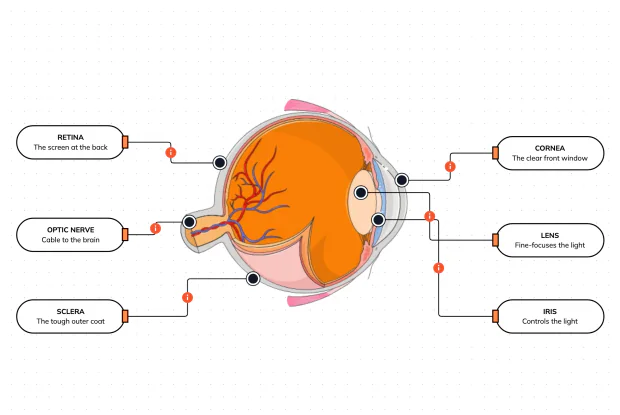
Your eye works like a living camera. Cut it open and label the cornea, lens, iris, retina, optic nerve and sclera, and follow light as it is focused onto a screen at the back of the eye and sent to the brain — in an interactive playground.