What Muscles Move Your Eyes?
Root Concept
Each eyeball is aimed by small strap-like muscles that run from the back of the bony eye socket to the surface of the eye. They work in opposing pairs — one rolls the eye up, its partner rolls it down, others turn it side to side — all anchored at a point deep in the socket, so the muscles can pull the eye to point your gaze anywhere.
CodePLU Goal
Upgrading Human Mental Models
Learn how to think in Workflows
Concept Development By codeplu.com
The muscles that move the eye, each joined to the job it does — rolling, aiming and anchoring the eyeball inside its bony socket
How Do Your Eyes Point Exactly Where You Want to Look?
Your eyes are almost never still. As you read this line they are flicking from word to word; glance up and they snap to something across the room; follow a bird and they track it smoothly across the sky. All of that aiming is done by muscles — small, strap-like muscles attached to each eyeball that pull it to point your gaze wherever you want, faster and more precisely than almost any other movement your body makes. You never feel them working, but they are among the busiest muscles you own.
Each eyeball sits in a bony cup in the skull called the eye socket, or orbit, and the muscles run from the deep back of that socket forward to attach onto the surface of the eyeball. When a muscle shortens, it tugs the eye and rotates it in its socket. There is one along the top that rolls the eye upward, one along the bottom that rolls it downward, and others on the sides that turn it left and right — six in all around each eye, together able to aim it in any direction.
Like muscles everywhere, they can only pull, never push, so they work in opposing pairs: to look up, the top muscle pulls while the bottom one relaxes; to look down, the reverse. All of them are rooted at a single point deep at the back of the socket, which gives them something firm to pull against. In the playground above you will label the eyeball, the muscles that move it up and down, their anchor point, and the bony socket that holds it all — and the label on each connection tells you the job that part does.
What Moves Your Eye — Part by Part?
What is actually being moved?
The thing being aimed is the eyeball itself — the whole round eye, sitting in its socket. When you 'move your eyes', you are not moving the coloured part or the pupil on its own; the entire eyeball swivels as one, rotating in place like a ball turning in a cup. Because it turns rather than slides, a small pull from a muscle attached near its surface can swing your gaze a long way. This is why your eyes can dart so quickly and land so precisely: they are lightweight balls, well lubricated, spinning on the spot, and the muscles only have to rotate them a little to point them at a completely different part of the world.
What are the muscles that move the eye?
Around each eyeball are six small muscles, called the extraocular ('outside-the-eye') muscles. Most of them are straight straps that run from the back of the socket forward to attach onto the eyeball — the figure shows two clearly, one along the top and one along the bottom. Each one, when it contracts, pulls the eye toward itself and so rotates your gaze in that direction: the top muscle rolls the eye up, the bottom one rolls it down, and the pair on the sides turn it left and right. Between them, these six muscles can aim the eye in any direction you like. They are unusually fast and precise muscles, because aiming your eyes accurately — and quickly — matters enormously for seeing clearly.
Why do they come in pairs?
Because a muscle can only pull, never push — the same rule you saw at the elbow and the knee. A single muscle could roll the eye up, but it could never push it back down again; for that you need a second muscle pulling the opposite way. So the eye muscles are arranged in opposing pairs: the top and bottom muscles are partners, and so are the two side muscles. To look up, the top muscle pulls while the bottom one relaxes and lengthens; to look down, the bottom pulls while the top relaxes. This push-me-pull-you teamwork, with one muscle of each pair always doing the opposite of the other, is how the eye can be aimed smoothly and held steady in any position rather than just flicking to one extreme.
Where are the muscles anchored?
For a muscle to pull the eye, its other end must be fixed to something solid — and for most of the eye muscles that fixed point is deep at the back of the socket, where they all take root together at a small ring of tissue attached to the bone. You can see the muscles in the figure converging to that point at the back. This gives them a firm anchor to pull against: when the muscle shortens, the anchored end stays put and the eyeball end is dragged toward it, rotating the eye. It is the same principle as every muscle in the body — one end anchored, the other end moving the part you want to move. Here the anchor is buried safely at the back of the bony socket, well out of harm's way.
What is the eye socket?
The eye socket, or orbit, is the deep bony cup in the skull that holds the eyeball, its muscles, and the nerves and blood vessels that serve them. Its job is protection and support: the surrounding bone shields the soft eye from knocks (the rim of the socket is what takes the blow if something hits your eye area), and it gives the muscles a solid frame to anchor to and pull against. You can feel the strong bony edge of your own orbit by pressing gently around your eye. Each eye has its own socket, angled slightly outward, and the eye sits cushioned in fat inside it so it can swivel freely while staying protected. Muscles need bone to work against, and the orbit is the bone the eye's muscles use.
Real World Example
Why Do Both of Your Eyes Always Move Together?
The eye muscles do not just move one eye; they keep both eyes perfectly coordinated so you see a single, steady world. A few everyday things show them working — and what happens when they don't:
Why you can't look with just one eye
The muscles working as a team
No matter how hard you try, you cannot point one eye left and the other right — both eyes always swing together in the same direction. That is because the brain never commands a single eye muscle alone; it moves the matching muscles of both eyes at once, so the two eyeballs stay aimed at the same point. This teamwork is essential: your two eyes see slightly different views, and the brain combines them into one three-dimensional picture — but only if they are pointed at exactly the same thing. If the eyes drifted independently, you would see double. So every glance is really twelve muscles, six per eye, being coordinated in perfect step by the brain.
Why your eyes jump as you read
Fast muscle flicks
Reading feels smooth, but your eyes are not gliding along the line — they are jumping. The eye muscles snap your gaze from one clump of words to the next in tiny, lightning-fast flicks, pausing briefly on each group to take it in, then flicking on. These jumps are among the fastest movements the human body can make, and they happen a few times every second as you read. You never notice because vision is briefly dialled down during each flick. It is the eye muscles' speed and precision on display: they can fire off an exact, aimed movement and stop it dead on target in a fraction of a second, hundreds of thousands of times a day.
What a squint is
Muscles out of balance
Sometimes the delicate balance between the eye muscles is off, and the two eyes no longer point at the same place — one may turn in, out, up or down relative to the other. This is a squint (also called strabismus, or being 'cross-eyed'), and it usually comes from the eye muscles of the two eyes not pulling in a coordinated way, whether because one is weaker or the brain's control is mismatched. Because the eyes are aimed differently, they send the brain two views that don't line up, which can cause double vision. It is why a squint is often treated by adjusting or strengthening the eye muscles — the problem is not the eye itself but the muscles aiming it. It shows, in reverse, just how finely those muscles normally keep both eyes locked together.
FAQs
Final Words
Your gaze is aimed by muscle. Each eyeball is swivelled in its bony socket by six small strap-like muscles that run from an anchor deep at the back of the socket to the surface of the eye. Because muscles can only pull, they work in opposing pairs — one rolls the eye up while its partner rolls it down, others turn it side to side — and the bony orbit both protects the eye and gives the muscles something firm to pull against. Name the eyeball, the up and down muscles, their anchor and the socket, and you understand how the eye is aimed.
These muscles are among the fastest and most coordinated in the body: they flick your eyes across a page, track a moving object, and keep both eyes locked on the same point so you see one clear world instead of two. With the eye seen from the outside, from the inside, and now the muscles that aim it, you have the whole eye. The head-to-toe tour of the body continues down from here — next, into the neck and spine.
Continue This Track
This concept is part 13 of The Human Body, Head to Toe.
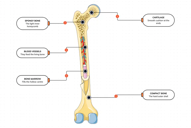
What Is a Bone Made Of?
Cut a bone open and it is nowhere near solid: a hard outer shell, a light honeycomb inside, a hollow centre full of marrow, blood vessels threading through, and a smooth cushion at each end. Learn what each part does — then label them yourself in an interactive playground.
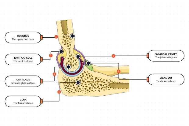
What Is Inside Your Elbow Joint?
A joint is where two bones meet and move — and it is far more than just the bones. Cut open the elbow and you find smooth cartilage caps, a sealed capsule of slippery fluid, and tough ligaments holding it all together. Learn what each part does, then label them yourself in an interactive playground.
What Makes Your Elbow Bend?
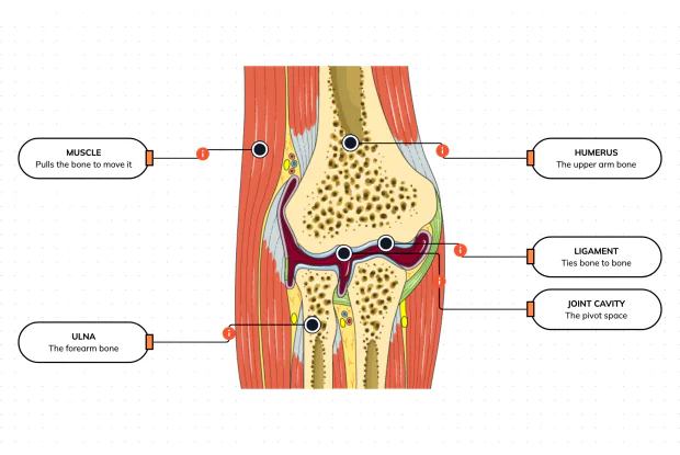
Bones and joints are just the hardware; nothing moves until a muscle pulls. Label the elbow to see the parts that turn a joint into movement — the muscle that pulls, the ligament that holds bone to bone, the two bones it links, and the space they pivot in — then build it yourself in an interactive playground.
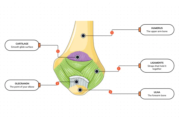
What Holds Your Elbow Together?
Your elbow is pulled on all day — by gravity, by every bag you carry, by every time you hang or push. So what stops the two bones from simply coming apart? Look at the back of the elbow and label the ligaments, the bony point, the cartilage and the two bones — and learn what holds the whole joint together.
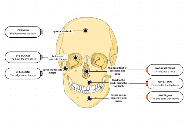
What Are the Main Bones of the Skull?
The skull looks like a single bone, but it is really about twenty-two locked together — with just one left free to move. Learn what each main part does, then label a skull yourself in an interactive playground.
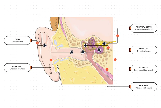
What Are the Parts of Your Ear?
Hearing is a chain of parts passing a sound along, each handing it to the next. Label the pinna, ear canal, eardrum, ossicles, cochlea and auditory nerve, and follow a sound all the way from the air outside to a signal your brain can read — in an interactive playground.
How Does Your Ear Keep You Balanced?
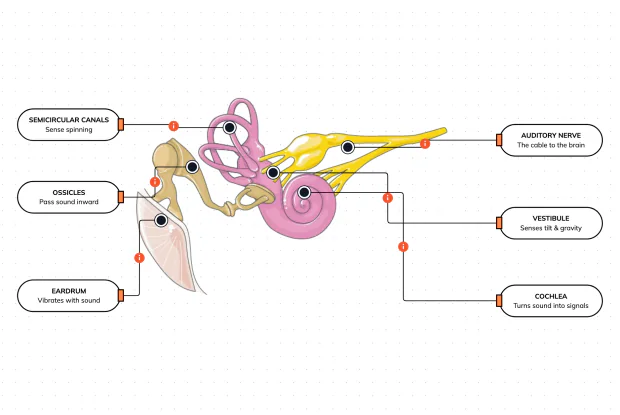
Your ear does a second, hidden job: balance. Inside it sit fluid-filled loops that sense spinning and a chamber that senses tilt and gravity. Label the semicircular canals, vestibule, cochlea, ossicles, eardrum and nerve, and learn how one organ both hears and keeps you upright — in an interactive playground.
What Are the Parts of the Eye You Can See?
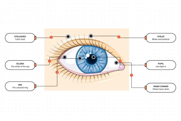
Before you look inside the eye, learn the parts you can already see in a mirror. Label the pupil, iris, sclera, eyelid, eyelashes and the tear-duct corner, and learn what each visible part does to let in light and protect the eye — in an interactive playground.
Where Do Your Tears Come From and Go?
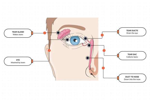
Your eyes make and drain tears all day to stay clean and wet. Label the tear gland, the eye, the tear ducts, the tear sac and the duct to the nose, and follow a tear from where it is made to where it drains — in an interactive playground.
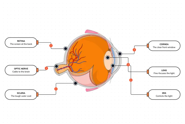
How Does Your Eye Turn Light Into a Picture?
Your eye works like a living camera. Cut it open and label the cornea, lens, iris, retina, optic nerve and sclera, and follow light as it is focused onto a screen at the back of the eye and sent to the brain — in an interactive playground.