What Is the Duodenum, the Gut's Mixing Bowl?
Root Concept
The duodenum is the short, C-shaped first part of the small intestine, curving around the head of the pancreas. Its upper end receives acidic, half-digested food from the stomach; bile from the liver and juice from the pancreas empty in to neutralise the acid and digest the food; its folded, muscular wall mixes everything; and its lower end passes the food on to the jejunum.
CodePLU Goal
Upgrading Human Mental Models
Learn how to think in Workflows

Concept Development By codeplu.com
The duodenum, each part joined to the job it does — receiving the stomach's acid, mixing in the digestive juices, and passing food on
What Happens the Moment Food Leaves Your Stomach?
When your stomach has finished churning a meal into a thick, sour soup, it does not release it all at once — it squirts it, a little at a time, into the very first stretch of the small intestine: the duodenum. This short, C-shaped tube, only about 25 centimetres long, is one of the busiest and most important places in your whole digestive system. It is where the real chemical digestion gets going, and where three different organs' outputs all come together at once. If the small intestine is where food is absorbed, the duodenum is the mixing bowl that gets it ready.
Its shape is distinctive: a tight C-curve that wraps neatly around the head of the pancreas — no coincidence, because the pancreas empties its digestive juice right into this curve. At the top, the duodenum begins where the stomach hands over its contents through a muscular valve. From there it sweeps down and around the pancreas, then straightens out and continues on to the rest of the small intestine at its far end.
What makes the duodenum special is what arrives here. The stomach's contents are strongly acidic — but the small intestine cannot cope with that acid, so the duodenum receives alkaline bile from the liver and gallbladder and alkaline juice from the pancreas, which together neutralise the acid and pour in the enzymes that break down fats, proteins and starches. The folded, muscular wall mixes it all thoroughly. In the playground above you will label the duodenum, its upper end, inner folds, wall and lower end — and the label on each connection tells you the job that part does.
What Does Each Part of the Duodenum Do?
What is the duodenum, and why is it C-shaped?
The duodenum is the first part of the small intestine — a short, wide tube, roughly 25 centimetres long, that connects the stomach to the rest of the gut. Its striking C-shape is functional: the curve wraps closely around the head of the pancreas, and that snug fit is exactly why the pancreas can empty its digestive juice straight into the duodenum, and why the bile duct from the liver joins at the same spot. So the C is not just a way to fit the tube in; it places the duodenum right up against the organs whose juices it needs. Its name even comes from its length — 'duodenum' derives from the Latin for twelve, because it is about twelve finger-widths long. Short, wide and curved, it is built to be a mixing chamber, not a long absorbing tube like the coils that follow.
What arrives at the upper end?
The upper end of the duodenum is where it joins the stomach, through a ring of muscle called the pyloric valve. The stomach spends hours churning a meal into a thick, acidic soup, and then releases it into the duodenum in small, controlled squirts through this valve — not all at once, but a little at a time, so the small intestine is never overwhelmed. What arrives is harsh: strongly acidic, only partly digested, and needing a lot of work before its nutrients can be absorbed. This is why the very start of the small intestine has to be a robust processing station rather than a delicate absorbing surface. Everything that follows in the duodenum is a response to what comes through this valve: acid to neutralise, and food to finish breaking down.
Why is this where bile and pancreatic juice enter?
Because this is exactly where they are needed. The acidic soup from the stomach would damage the rest of the small intestine and would stop the digestive enzymes working, so it has to be dealt with immediately — and the duodenum is where both the liver and the pancreas deliver their juices to do the job. Bile from the liver and gallbladder, and juice from the pancreas, both flow in through a shared opening into the duodenum's curve. These juices are alkaline, so they neutralise the stomach's acid, calming the soup down; and they are full of enzymes and bile salts that break down fats, proteins and starches. So the duodenum is a genuine crossroads: the stomach, the liver and the pancreas all deliver here, and the duodenum is where their contents are combined. It is the single busiest junction in digestion.
What do the inner folds and wall do?
Like the rest of the small intestine, the duodenum's inner lining is thrown into folds and covered in tiny projections, giving it a large surface — so absorption of some nutrients (and minerals like iron and calcium) begins right here. But the duodenum's lining has an extra challenge: it is the first tissue to meet the stomach's acid, so it also releases mucus and signals to help protect itself and to trigger the neutralising juices. The muscular wall, meanwhile, does the mixing: it churns the incoming food together with the bile and pancreatic juice, so the enzymes reach every part of the soup, and then moves the mixture steadily onward. Folds to absorb and protect, muscle to mix and move — the duodenum's wall is a workbench where the meal is thoroughly prepared for the long absorbing journey ahead.
Where does the food go next?
At its lower end, the duodenum straightens out and continues seamlessly into the next part of the small intestine, the jejunum. By this point the food has been transformed: the acid is neutralised, the fats are broken into droplets by bile, and the pancreatic enzymes have begun dismantling the proteins and starches. In other words, the hard preparation is done, and what passes onward is a well-mixed, chemically-primed liquid ready for the main event — absorption along the metres of coiled jejunum and ileum that follow. So the duodenum is best thought of as the setup stretch: it receives, neutralises, mixes and starts the digestion, then hands a properly-prepared meal on to the long absorbing tube. Everything downstream depends on the work done in this short first curve.
Real World Example
Why Do So Many Ulcers Form Just Past the Stomach?
The duodenum is where the acidic stomach contents meet the alkaline juices of the liver and pancreas. Following that hand-over explains both how digestion is set up and why this spot is prone to trouble:
The acid bath arrives
The upper end
The stomach delivers its contents to the duodenum as a thick, strongly acidic soup — acid it made deliberately, to kill germs and start breaking down proteins. But that acid is a problem the moment it leaves the stomach: the stomach has a thick, specialised lining built to withstand it, whereas the small intestine does not. The duodenum receives this acid bath first, in controlled squirts through the pyloric valve, and must deal with it immediately before the delicate absorbing tube downstream is exposed. So the very start of the small intestine is, in effect, on the front line — the first tissue in the gut to face the full strength of stomach acid every time you eat. That front-line position is the key to everything that follows, good and bad.
The great neutralising
Bile and pancreatic juice
To handle the incoming acid, the duodenum calls in reinforcements: bile from the liver and gallbladder, and juice from the pancreas, both of which are alkaline and both of which empty into the duodenum's curve. As they mix with the acidic soup, they neutralise it — bringing it from harshly acidic to gently alkaline, the conditions the intestine's own enzymes need to work. At the same time these juices flood in the enzymes and bile salts that finish breaking fats, proteins and starches into absorbable pieces. This is the duodenum's masterstroke: it does not just survive the acid, it converts a hostile acid bath into a friendly, enzyme-rich soup, all within a few centimetres. The whole rest of digestion depends on this neutralising step happening reliably, meal after meal.
Why ulcers form here
The lining under attack
Because the duodenum meets the acid first, it is also the most common place in the gut for an ulcer — a raw, eroded sore in the lining. Normally the balance holds: mucus protects the lining and the alkaline juices neutralise the acid fast enough. But if too much acid arrives, or the protective mucus is weakened (often by an infection with a particular bacterium, or by certain painkillers), the acid can eat into the duodenal lining and form a duodenal ulcer. This is why 'stomach ulcers' are in fact frequently just past the stomach, in the duodenum, and why they often hurt between meals when acid sits unneutralised. It is a direct consequence of the duodenum's role: the tissue that faces the acid first is the tissue most at risk when the defences fail.
FAQs
Final Words
The duodenum is short but pivotal — the C-shaped first stretch of the small intestine and the busiest crossroads in digestion. Its upper end receives the stomach's acidic soup through a valve; wrapped around the pancreas, it is where bile from the liver and juice from the pancreas empty in to neutralise that acid and pour in enzymes; its folded, muscular wall mixes everything thoroughly; and its lower end passes a well-prepared meal on to the jejunum. Receive, neutralise, mix, hand on — that is the duodenum's job in four steps.
Its front-line position also explains why duodenal ulcers are so common: it is the first tissue to face the stomach's acid every time you eat. With the small intestine's overall design and now its critical first stretch understood, the natural next zoom is inward — to the tiny villi carpeting the lining, where an absorbed nutrient finally crosses from the gut into your blood.
Continue This Track
This concept is part 36 of The Human Body, Head to Toe.
What Is a Bone Made Of?
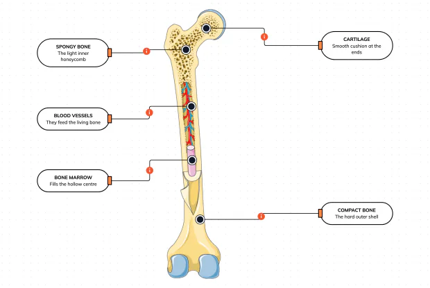
Cut a bone open and it is nowhere near solid: a hard outer shell, a light honeycomb inside, a hollow centre full of marrow, blood vessels threading through, and a smooth cushion at each end. Learn what each part does — then label them yourself in an interactive playground.
What Is Inside Your Elbow Joint?
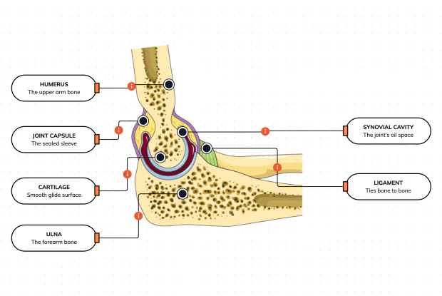
A joint is where two bones meet and move — and it is far more than just the bones. Cut open the elbow and you find smooth cartilage caps, a sealed capsule of slippery fluid, and tough ligaments holding it all together. Learn what each part does, then label them yourself in an interactive playground.
What Makes Your Elbow Bend?
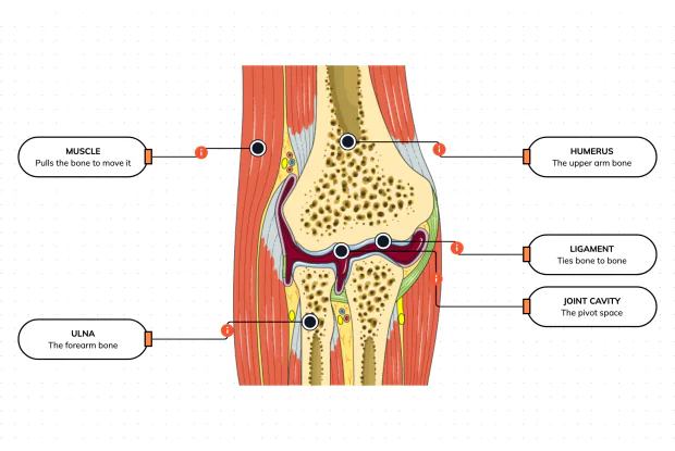
Bones and joints are just the hardware; nothing moves until a muscle pulls. Label the elbow to see the parts that turn a joint into movement — the muscle that pulls, the ligament that holds bone to bone, the two bones it links, and the space they pivot in — then build it yourself in an interactive playground.
What Holds Your Elbow Together?
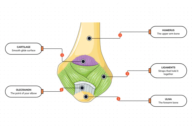
Your elbow is pulled on all day — by gravity, by every bag you carry, by every time you hang or push. So what stops the two bones from simply coming apart? Look at the back of the elbow and label the ligaments, the bony point, the cartilage and the two bones — and learn what holds the whole joint together.
What Are the Main Bones of the Skull?
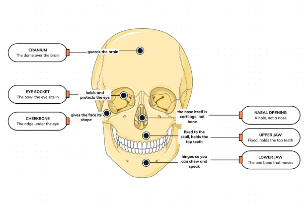
The skull looks like a single bone, but it is really about twenty-two locked together — with just one left free to move. Learn what each main part does, then label a skull yourself in an interactive playground.
What Are the Parts of Your Ear?
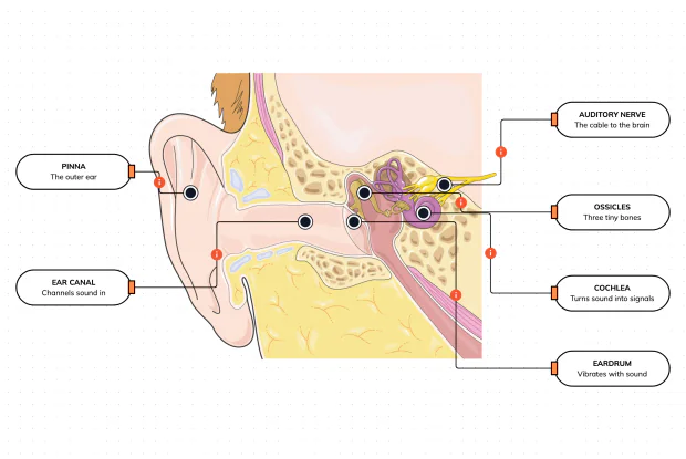
Hearing is a chain of parts passing a sound along, each handing it to the next. Label the pinna, ear canal, eardrum, ossicles, cochlea and auditory nerve, and follow a sound all the way from the air outside to a signal your brain can read — in an interactive playground.
How Does Your Ear Keep You Balanced?
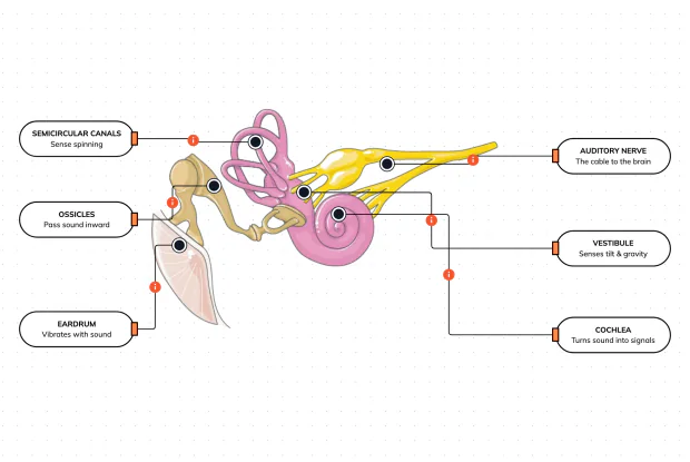
Your ear does a second, hidden job: balance. Inside it sit fluid-filled loops that sense spinning and a chamber that senses tilt and gravity. Label the semicircular canals, vestibule, cochlea, ossicles, eardrum and nerve, and learn how one organ both hears and keeps you upright — in an interactive playground.
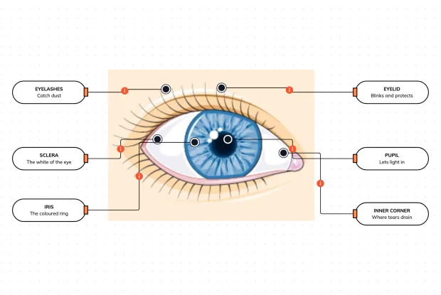
What Are the Parts of the Eye You Can See?
Before you look inside the eye, learn the parts you can already see in a mirror. Label the pupil, iris, sclera, eyelid, eyelashes and the tear-duct corner, and learn what each visible part does to let in light and protect the eye — in an interactive playground.
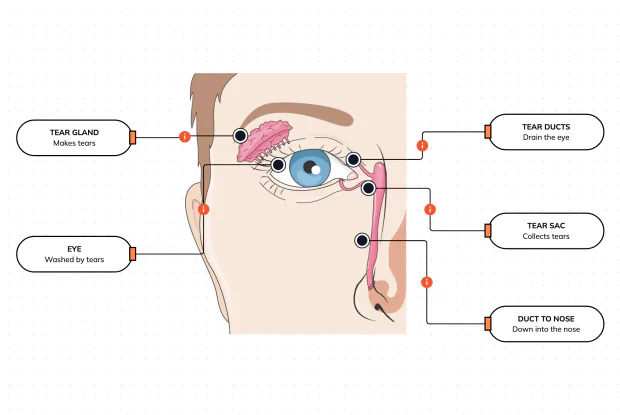
Where Do Your Tears Come From and Go?
Your eyes make and drain tears all day to stay clean and wet. Label the tear gland, the eye, the tear ducts, the tear sac and the duct to the nose, and follow a tear from where it is made to where it drains — in an interactive playground.
How Does Your Eye Turn Light Into a Picture?
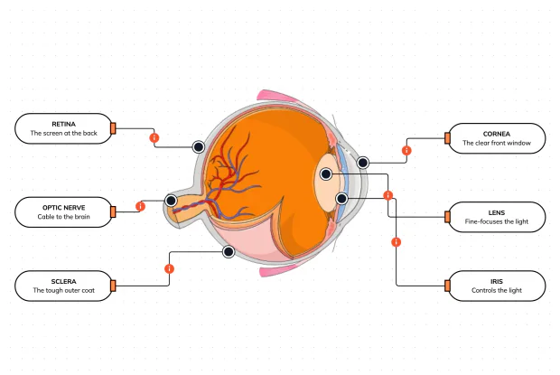
Your eye works like a living camera. Cut it open and label the cornea, lens, iris, retina, optic nerve and sclera, and follow light as it is focused onto a screen at the back of the eye and sent to the brain — in an interactive playground.