What Are the Bones of the Leg?
Root Concept
The leg has one bone in the thigh (the femur, the body's longest and strongest), a kneecap in front (the patella), and two below the knee — the thick weight-bearing tibia and the slim fibula — meeting the foot at the ankle hinge.
CodePLU Goal
Upgrading Human Mental Models
Learn how to think in Workflows

Concept Development By codeplu.com
Each bone of the leg, joined to the job its shape is built for — bear weight, protect, or steady
Illustration: AI-generated for CodePLU
Why Does the Leg Have One Bone Up Top and Two Below the Knee?
Stand up and press a hand into the front of your thigh, then slide it down past your knee to your shin. Above the knee you can feel one big bone; below it, if you press on the outer side, you can feel a second, thinner one. That is the leg's basic plan: a single thick bone in the thigh, and a pair of bones in the lower leg — the very same one-then-two pattern the arm uses, but built for a heavier job.
The single thigh bone, the femur, is the longest and strongest bone in your whole body, because it has the hardest task: carrying your entire weight and swinging the leg from the hip with every step. Below the knee the work splits between two bones. The thick tibia — your shin bone — takes almost all of the weight, standing directly under the femur, while the slim fibula alongside it carries hardly any load and instead anchors muscles and steadies the ankle. In front of the knee sits a small extra bone, the kneecap or patella, protecting the joint and helping the thigh muscle straighten the leg.
Where the tibia and fibula reach the foot they form the ankle, a hinge that lets the foot tip up and down. In the playground above you will label the femur, the patella, the tibia, the fibula and the ankle — and the label on each connection tells you the job that part does.
What Does Each Bone of the Leg Do?
Why is the femur the biggest bone in the body?
The femur is the single bone of the thigh, running from the hip down to the knee, and it is the longest and strongest bone you have. It has to be: it carries your entire body weight and acts as the long lever that swings your leg forward from the hip with every stride. A bone that both bears that load and takes the force of running and jumping has to be thick and solid, so the femur is built heavier than anything else in the skeleton. Its rounded head plugs into the deep hip socket, and its wide lower end forms the top half of the knee.
What is the patella, and why is it there?
The patella is the kneecap — a small, roughly triangular bone that sits in the tendon crossing the front of the knee. It does two jobs. First, it shields the knee joint: it is the bone you land on when you fall forward onto your knees, taking the knock instead of the softer joint behind it. Second, it works like a pulley. The big thigh muscle pulls on the shin through that tendon, and the patella lifts the tendon away from the joint, giving the muscle more leverage to straighten the leg — which is what powers you up from a squat or a stair.
Why does the tibia carry the weight and the fibula not?
Below the knee the leg has two bones, but they share the load very unevenly. The tibia — the shin bone — is the thick one, and it sits directly beneath the femur, so your weight passes straight down through it to the ankle. It is the bone you can feel as the hard ridge down the front of your lower leg. The fibula is the slim bone on the outer side, and it barely bears weight at all. Instead it serves as an anchor for muscles and, at its lower end, forms the outer knob of the ankle that stops the joint rolling too far. Two bones, but only one is the pillar.
What holds the tibia and fibula together?
The tibia and fibula do not just lie side by side — they are lashed together along their whole length by a tough sheet of tissue between them, and joined more firmly at the top, just below the knee, and at the bottom, at the ankle. That binding keeps the pair working as a unit and stops them splaying apart when you land. It also means the two bones share and spread sudden forces: a hard twist that might otherwise snap one bone alone is partly passed to the other and to the connecting sheet.
What is the ankle made of?
The ankle is the joint where the two lower-leg bones meet the foot. The tibia and fibula form a squared-off socket — the tibia making the inner knob and the roof, the fibula making the outer knob — and the top bone of the foot, the talus, sits snugly inside it. That grip is what makes the ankle a hinge: it lets the foot tip up and down for walking, while the knobs on each side stop it sliding sideways. It is the handover point where this tutorial passes on to the bones of the foot.
Real World Example
Why Can a Broken Fibula Sometimes Still Let You Walk, but a Broken Tibia Can't?
Footballers and hikers sometimes crack the slim outer bone of the lower leg and, to everyone's surprise, can still hobble on it — while a broken shin bone puts you flat on the ground. The difference is not luck; it is which bone does the load-bearing:
The tibia is the pillar
It carries the weight
The tibia sits directly under the femur, in a straight line from your knee to your ankle, so your entire body weight travels down through it every time you stand on that leg. It is a structural pillar, and like any pillar it only works while it is whole. Break the tibia and that line of support is severed: the moment you try to put weight on the leg, the broken ends grind and give way, which is why a tibia fracture drops you and normally needs a cast or surgery and weeks off the leg. There is no other bone positioned to take over its load-bearing job.
The fibula is a strut, not a pillar
It barely bears weight
The fibula runs alongside the tibia on the outer side, but it is thin and set off to the edge, well away from the straight line your weight travels down. Its real jobs are to anchor muscles and to form the outer knob of the ankle, not to hold you up — it carries only a small fraction of your weight. So when the fibula alone cracks, the tibia is still an intact pillar underneath you, and the leg can often still bear weight enough to limp. It hurts, and it still needs to heal, but the support structure has not been broken.
What this tells you
Position decides the job
Two bones can sit inches apart and yet matter completely differently, and the reason is where each one stands relative to the load. The tibia is on the line of force, so it is thick, central and indispensable; the fibula is off to the side, so it can be slim and, in a pinch, spared. This is the same lesson the whole skeleton keeps teaching: a bone's shape and importance follow directly from the job its position gives it. Read where a bone sits and you can predict how much it carries — and how badly you will miss it if it breaks.
FAQs
Final Words
The leg is the arm's plan rebuilt for weight. One thick femur in the thigh — the body's longest, strongest bone — carries the load and swings the limb from the hip; a kneecap rides in front to protect the joint and give the thigh muscle leverage; and below the knee the job splits between a thick weight-bearing tibia and a slim fibula that steadies rather than carries. Nothing is arbitrary: each bone's size and place follow from the job it has to do.
Label the femur, the patella, the tibia, the fibula and the ankle, and the next time you climb a stair or feel the hard ridge of your shin you will know exactly which bone is doing the work. Where the tibia and fibula meet the foot, the skeleton hands over to the bones of the foot — the last step of the journey down the body.
Continue This Track
This concept is part 20 of The Human Body, Head to Toe.
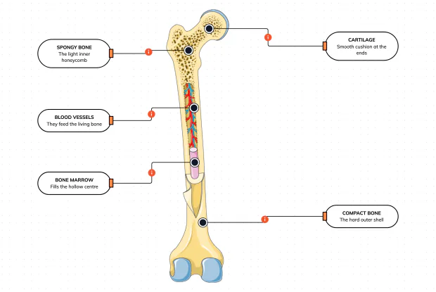
What Is a Bone Made Of?
Cut a bone open and it is nowhere near solid: a hard outer shell, a light honeycomb inside, a hollow centre full of marrow, blood vessels threading through, and a smooth cushion at each end. Learn what each part does — then label them yourself in an interactive playground.
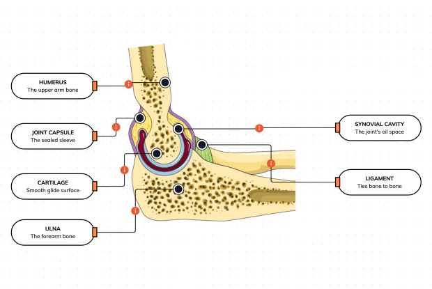
What Is Inside Your Elbow Joint?
A joint is where two bones meet and move — and it is far more than just the bones. Cut open the elbow and you find smooth cartilage caps, a sealed capsule of slippery fluid, and tough ligaments holding it all together. Learn what each part does, then label them yourself in an interactive playground.
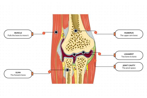
What Makes Your Elbow Bend?
Bones and joints are just the hardware; nothing moves until a muscle pulls. Label the elbow to see the parts that turn a joint into movement — the muscle that pulls, the ligament that holds bone to bone, the two bones it links, and the space they pivot in — then build it yourself in an interactive playground.
What Holds Your Elbow Together?
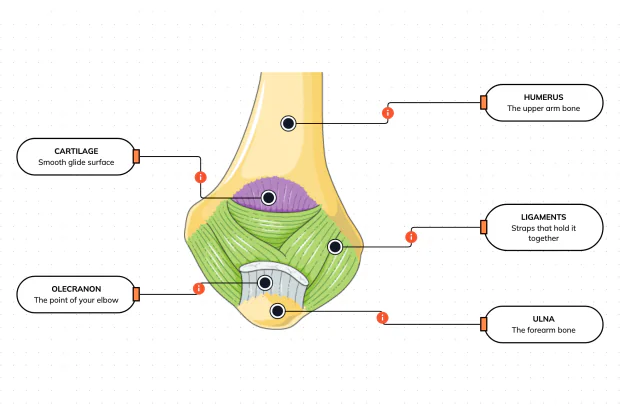
Your elbow is pulled on all day — by gravity, by every bag you carry, by every time you hang or push. So what stops the two bones from simply coming apart? Look at the back of the elbow and label the ligaments, the bony point, the cartilage and the two bones — and learn what holds the whole joint together.
What Are the Main Bones of the Skull?
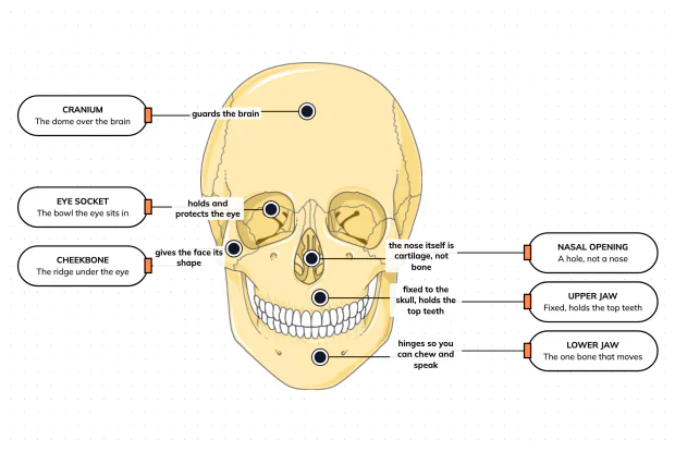
The skull looks like a single bone, but it is really about twenty-two locked together — with just one left free to move. Learn what each main part does, then label a skull yourself in an interactive playground.
What Are the Parts of Your Ear?
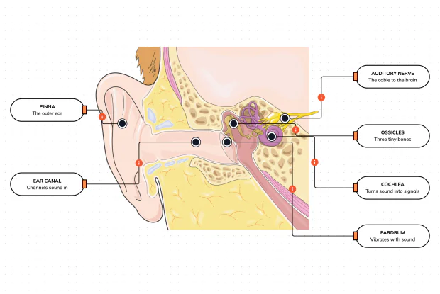
Hearing is a chain of parts passing a sound along, each handing it to the next. Label the pinna, ear canal, eardrum, ossicles, cochlea and auditory nerve, and follow a sound all the way from the air outside to a signal your brain can read — in an interactive playground.
How Does Your Ear Keep You Balanced?
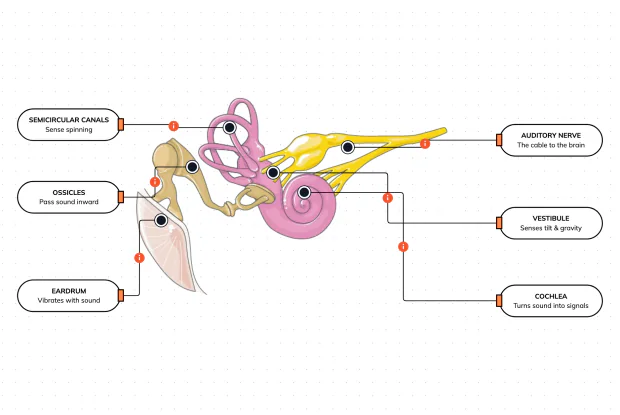
Your ear does a second, hidden job: balance. Inside it sit fluid-filled loops that sense spinning and a chamber that senses tilt and gravity. Label the semicircular canals, vestibule, cochlea, ossicles, eardrum and nerve, and learn how one organ both hears and keeps you upright — in an interactive playground.
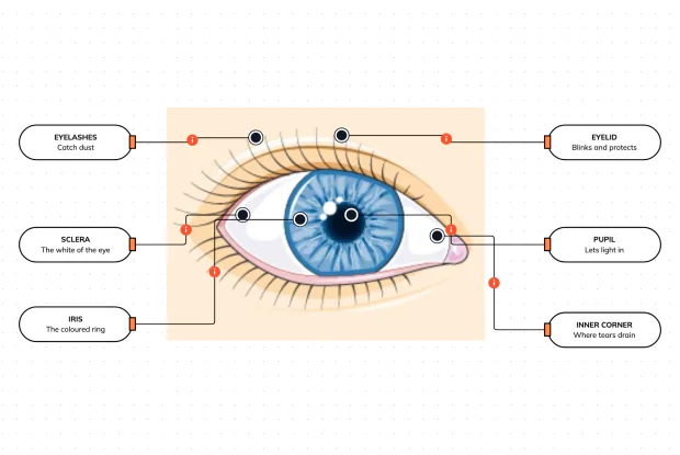
What Are the Parts of the Eye You Can See?
Before you look inside the eye, learn the parts you can already see in a mirror. Label the pupil, iris, sclera, eyelid, eyelashes and the tear-duct corner, and learn what each visible part does to let in light and protect the eye — in an interactive playground.
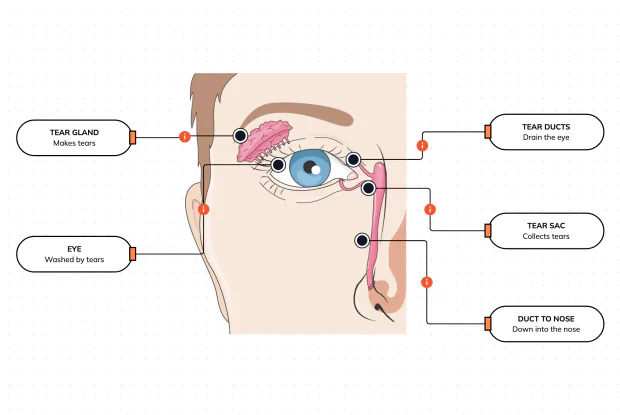
Where Do Your Tears Come From and Go?
Your eyes make and drain tears all day to stay clean and wet. Label the tear gland, the eye, the tear ducts, the tear sac and the duct to the nose, and follow a tear from where it is made to where it drains — in an interactive playground.
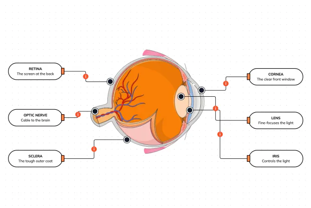
How Does Your Eye Turn Light Into a Picture?
Your eye works like a living camera. Cut it open and label the cornea, lens, iris, retina, optic nerve and sclera, and follow light as it is focused onto a screen at the back of the eye and sent to the brain — in an interactive playground.