How Does Your Bladder Store Urine?
Root Concept
The bladder is a stretchy, muscular bag that stores urine. Two ureters bring urine in through ureter openings; folds called rugae let the muscular wall stretch as it fills; and when you are ready, the wall squeezes the urine out through the urethra.
CodePLU Goal
Upgrading Human Mental Models
Learn how to think in Workflows
Concept Development By codeplu.com
The bladder cut open — the ureters and their openings that fill it, the muscular wall and its stretchy folds, and the urethra that empties it
Where Does Your Pee Wait Until You're Ready?
Your kidneys make urine all the time, in a slow steady trickle, day and night. But you do not go to the toilet all the time — you go every few hours. So there must be somewhere for the urine to wait in between. That somewhere is your bladder: a stretchy, muscular bag that stores urine until you are ready to let it out.
The bladder sits low in your tummy. Urine reaches it through two tubes called the ureters, one coming down from each kidney, which empty into the bladder through small ureter openings. As more urine arrives, the bladder slowly fills and swells like a balloon, because its wall is made of muscle that can stretch a long way and springs back afterwards.
When the bladder is full enough, it sends a message to your brain — the feeling of needing to go. When you decide it is time, the muscular wall squeezes and pushes the urine out through a tube at the bottom called the urethra. In the playground you will label the ureters and their openings, the muscular wall, the folds that let it stretch, and the urethra.
How Does the Bladder Work?
How urine gets in
Urine does not appear in the bladder from nowhere — it is delivered. Each kidney makes urine and drips it into a long tube called a ureter. You have two ureters, one from the left kidney and one from the right, and they run down and enter the bladder near its base. Where each ureter joins, there is a small hole called a ureter opening, and urine trickles in through these two openings all day long. The openings are cleverly built so urine can flow in but not back up toward the kidneys, even when the bladder is full and under pressure. So the filling of the bladder is quiet and one-way: kidney makes it, ureter carries it, the ureter opening lets it in, drop by drop.
How the bladder stretches and stores
The bladder's job is to hold a growing amount of urine without leaking, and its wall is built for exactly that. The wall is made of muscle that can stretch to many times its empty size, so an empty bladder is small and wrinkled while a full one is round and smooth. The secret to the stretching is on the inside: when empty, the lining is gathered into folds called rugae, like the pleats in a folded paper fan. As urine fills the bladder, these folds flatten out and open up, letting the bag swell without tearing. This is why you can comfortably store quite a lot of urine between toilet trips — the muscular wall and its clever folds let the bladder grow to fit, then shrink back down once it is emptied.
How urine gets out
When the bladder has filled to a certain point, the stretch in its wall triggers a signal to your brain: the familiar feeling that you need to go. You can hold on for a while, because a ring of muscle at the bladder's exit stays squeezed shut like a drawstring. When you decide it is time, that ring relaxes and the muscular bladder wall squeezes inward, pushing the urine out through the urethra — the tube that leads from the bladder to the outside of the body. Once the bladder is empty, the wall relaxes, the folds gather back up, and the whole cycle begins again as fresh urine arrives from the kidneys. Filling is automatic; emptying is something you learn to control.
Real World Example
When Do You Notice Your Bladder?
The bladder is one organ you definitely notice several times a day. Here are three everyday moments:
The feeling of needing to go
A full bladder signals your brain
That growing feeling that you need the toilet is your bladder talking to your brain. As urine fills the bladder, its muscular wall stretches, and stretch sensors in the wall send a message up your nerves: 'getting full'. A little full, and you get a gentle reminder you can easily ignore; very full, and the message becomes urgent and hard to think past. This is a clever early-warning system — it lets you know in good time so you can find a toilet before the bladder is completely full. So the need-to-go feeling is not the urine itself; it is the stretch of the bladder wall being reported to your brain, getting louder as the bag fills up.
Holding it until you find a toilet
A ring of muscle keeps the exit shut
Being able to 'hold it' is thanks to a ring of muscle around the bladder's exit, where the urethra begins. Most of the time this ring stays squeezed shut, like a drawstring on a bag, keeping urine safely inside even as the bladder fills and presses. When you reach a toilet and decide it is time, your brain lets that ring relax and, at the same time, tells the bladder wall to squeeze — and out the urine flows through the urethra. Being able to keep the ring shut until the right moment is what lets you wait through a lesson or a car journey. It is a small muscle doing a very appreciated job.
Learning bladder control as you grow
Toilet training is learning to control the exit
Very young babies do not control their bladders at all — the moment it fills enough, it empties on its own, which is why babies wear nappies. As a child's brain and nerves develop, they gradually learn to sense the bladder filling and to hold the exit ring shut until they reach a toilet. That is what toilet training is: learning conscious control over a system that used to run entirely by itself. It also explains why some children wet the bed at night for a while — during deep sleep, the brain has not yet learned to notice the 'full' signal and keep the ring shut. Bladder control is a skill the body grows into, not something we are born with.
FAQs
Final Words
The bladder is a stretchy, muscular bag that stores your urine. Two ureters bring urine down from the kidneys and empty it in through ureter openings; folds called rugae let the muscular wall stretch as the bag fills; and when you are ready, the wall squeezes the urine out through the urethra. Filling happens automatically, drop by drop, while emptying is something you control.
That control is a skill you grew into — from nappies, to toilet training, to easily holding on until you find a toilet — all thanks to a small ring of muscle guarding the exit. Together with the kidneys and ureters, the bladder completes the urinary system: blood is cleaned in the kidneys, urine travels down the ureters, waits in the bladder, and finally leaves through the urethra.
Continue This Track
This concept is part 48 of The Human Body, Head to Toe.
What Is a Bone Made Of?
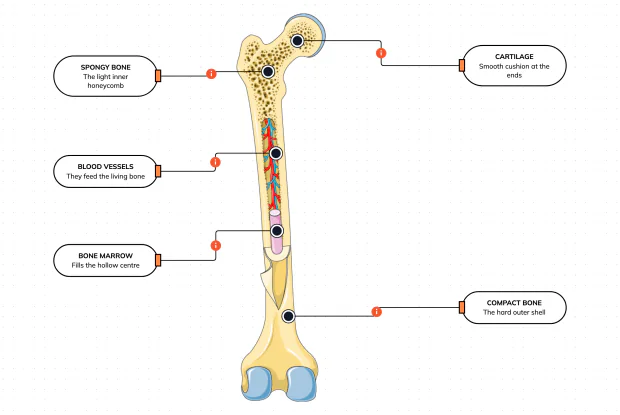
Cut a bone open and it is nowhere near solid: a hard outer shell, a light honeycomb inside, a hollow centre full of marrow, blood vessels threading through, and a smooth cushion at each end. Learn what each part does — then label them yourself in an interactive playground.
What Is Inside Your Elbow Joint?
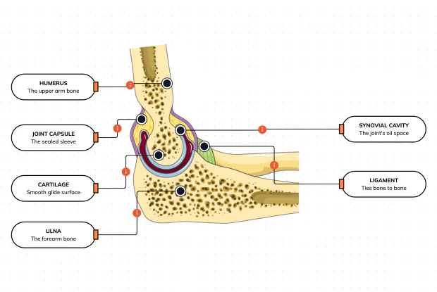
A joint is where two bones meet and move — and it is far more than just the bones. Cut open the elbow and you find smooth cartilage caps, a sealed capsule of slippery fluid, and tough ligaments holding it all together. Learn what each part does, then label them yourself in an interactive playground.
What Makes Your Elbow Bend?
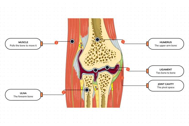
Bones and joints are just the hardware; nothing moves until a muscle pulls. Label the elbow to see the parts that turn a joint into movement — the muscle that pulls, the ligament that holds bone to bone, the two bones it links, and the space they pivot in — then build it yourself in an interactive playground.
What Holds Your Elbow Together?
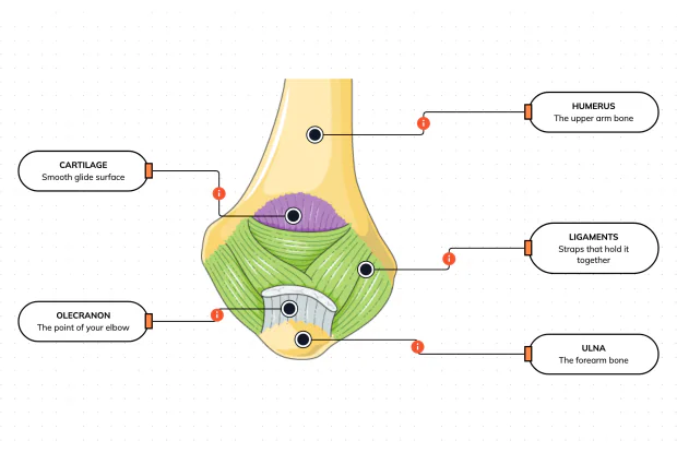
Your elbow is pulled on all day — by gravity, by every bag you carry, by every time you hang or push. So what stops the two bones from simply coming apart? Look at the back of the elbow and label the ligaments, the bony point, the cartilage and the two bones — and learn what holds the whole joint together.
What Are the Main Bones of the Skull?
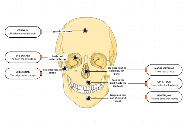
The skull looks like a single bone, but it is really about twenty-two locked together — with just one left free to move. Learn what each main part does, then label a skull yourself in an interactive playground.
What Are the Parts of Your Ear?
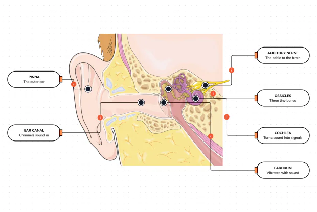
Hearing is a chain of parts passing a sound along, each handing it to the next. Label the pinna, ear canal, eardrum, ossicles, cochlea and auditory nerve, and follow a sound all the way from the air outside to a signal your brain can read — in an interactive playground.
How Does Your Ear Keep You Balanced?
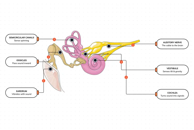
Your ear does a second, hidden job: balance. Inside it sit fluid-filled loops that sense spinning and a chamber that senses tilt and gravity. Label the semicircular canals, vestibule, cochlea, ossicles, eardrum and nerve, and learn how one organ both hears and keeps you upright — in an interactive playground.
What Are the Parts of the Eye You Can See?
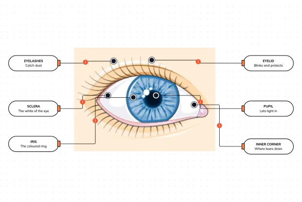
Before you look inside the eye, learn the parts you can already see in a mirror. Label the pupil, iris, sclera, eyelid, eyelashes and the tear-duct corner, and learn what each visible part does to let in light and protect the eye — in an interactive playground.
Where Do Your Tears Come From and Go?
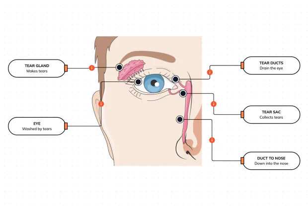
Your eyes make and drain tears all day to stay clean and wet. Label the tear gland, the eye, the tear ducts, the tear sac and the duct to the nose, and follow a tear from where it is made to where it drains — in an interactive playground.
How Does Your Eye Turn Light Into a Picture?
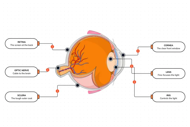
Your eye works like a living camera. Cut it open and label the cornea, lens, iris, retina, optic nerve and sclera, and follow light as it is focused onto a screen at the back of the eye and sent to the brain — in an interactive playground.