What Is a Cell, and What Does Each Part Do?
Root Concept
A cell is a working unit whose separate parts each carry out a different job.
CodePLU Goal
Upgrading Human Mental Models
Learn how to think in Workflows

Concept Development By codeplu.com
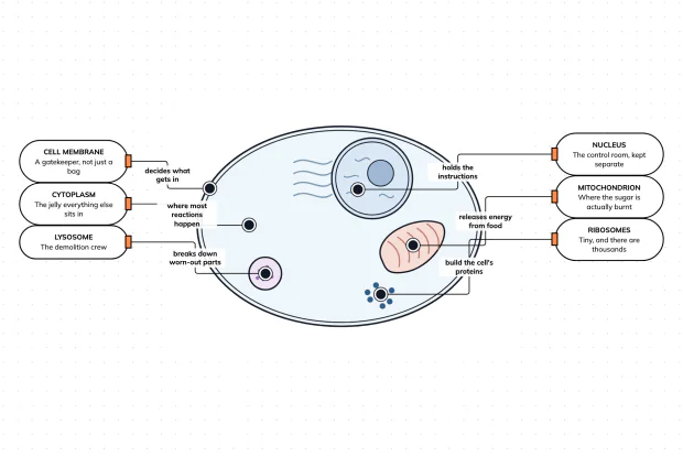
Each part of a cell joined to the job it does. The drawing is schematic and not to scale — a real ribosome is far smaller than this.
Zooming All the Way In
Biology so far has looked at whole things: a plant with roots and leaves, a food chain running from the sun through one living thing to the next. This tutorial goes the other way and zooms in until there is nothing smaller left that is still alive.
What you find is the cell. Every living thing is built from them — you are made of roughly thirty trillion, an oak tree of far more, and a bacterium is a single one. That is the first big idea in biology, and it took microscopes to find: nobody knew cells existed until Robert Hooke looked at a sliver of cork in 1665 and saw it was divided into tiny boxes, which he named after the small rooms monks lived in.
The second big idea is the one this tutorial is really about. A cell is not a bag of jelly with a dot in the middle. It is organised. Different jobs happen in different places inside it, each handled by a separate structure called an organelle, in much the same way that a hospital keeps its operating theatres, its kitchens and its records in separate rooms. In the playground below, six of those parts are waiting to be matched to the job each one does.
Six Jobs, and Why They Are Kept Apart
Why is a cell not just a bag of jelly?
Because the reactions that keep it alive would get in each other's way. A cell is building proteins, releasing energy, storing waste and destroying worn-out parts all at the same time, and some of those processes would ruin the others if they met. The solution is the same one a hospital uses: give each job its own room. Those rooms are the organelles, and most of them are wrapped in their own membrane so that what happens inside stays inside. The jelly itself has a name too — the cytoplasm — and it is not just filler. It is a crowded solution of water, salts and proteins, and the majority of the cell's chemical reactions happen in it rather than in any organelle.
What is the nucleus for, and why is it shut away?
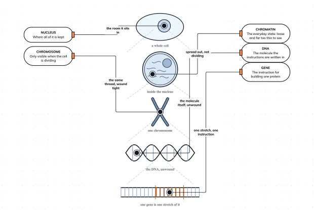
The nucleus holds the cell's instructions, written in DNA. Those instructions say how to build every protein the cell needs, and a protein is how almost everything in biology actually gets done. Inside the nucleus the DNA is wound into long threads called chromatin, and the darker blob you can see in the drawing is the nucleolus, where the parts of ribosomes are made. What matters is that the nucleus has its own double membrane around it. The instructions are the one thing a cell cannot replace if they are damaged, so they are kept behind a wall with controlled openings, and copies are sent out rather than the original. A cell that loses its nucleus can keep going for a while, but it cannot build anything new.
Where does a cell get its energy?
From the mitochondria, which is why they are usually described as the cell's power plants. Food brings in glucose, and a mitochondrion releases the energy stored in it through respiration, packaging that energy into a form the rest of the cell can spend. The folds you can see inside the bean shape are there for the same reason a radiator has fins: they pack a large working surface into a small space, and the reactions happen on that surface. The number of mitochondria is a good clue to what a cell does for a living. A muscle cell may hold thousands, because contracting is expensive. A cell that mostly sits still holds far fewer.
What builds things, and what clears them away?
Two organelles with opposite jobs, and it is worth meeting them as a pair. Ribosomes build proteins, following the instructions copied out of the nucleus. They are the small solid dots in the drawing, and they are the one part here that has no membrane around it — some float free in the cytoplasm and some sit on the folded sheets beside the nucleus. Lysosomes do the reverse. Each is a bag of digestive enzymes that breaks down worn-out organelles and anything the cell has swallowed, so the raw materials can be used again. Keeping those enzymes sealed inside a membrane matters: loose in the cytoplasm they would digest the cell itself.
Why is the membrane the part that matters most?
Because it decides what the cell is. Everything the cell needs has to cross it, and everything the cell must keep out is stopped by it, so the membrane is not a wall and not a simple bag — it is a gatekeeper that lets some substances through and refuses others. The proper word for that is selectively permeable, and it is the single most consequential property in this tutorial. It explains why a cell in pure water swells, why a cell in salty water shrinks, and why some substances cost the cell energy to take in while others drift in for free. The next two tutorials in this track are about nothing else.
Real World Example
Looking at Your Own Cheek Cells
The standard school practical is to scrape the inside of your cheek, put it on a slide and look. What you see is a useful shock, because it is nothing like the picture in the book.
Step 1
Almost nothing is visible
Under an ordinary school microscope a cheek cell is a pale, irregular smudge. You can usually make out its edge, which is the cell membrane, and a slightly darker spot near the middle, which is the nucleus. That is often all. The mitochondria, the ribosomes and the lysosomes are there in their thousands and none of them can be seen, because a light microscope cannot resolve anything much smaller than about two tenths of a micrometre and most organelles sit below that line. The parts are not missing from the cell; they are missing from the image.
Step 2
A stain makes the nucleus appear
Adding a drop of methylene blue or iodine changes the view completely. The dye is taken up unevenly, and it concentrates in the nucleus, which jumps out as a clear dark circle against a much paler cytoplasm. This is why staining is in every practical: the problem with a cheek cell is not that it is too small to see but that it is too transparent, and a stain fixes contrast rather than magnification. It is also a reminder that the colours in a textbook diagram are a teaching choice. Cells are not pink and blue.
Step 3
The diagram is a map, not a photograph
Everything beyond the nucleus and the membrane was discovered with the electron microscope, which arrived in the 1930s and sees structures a thousand times finer. The drawing in this tutorial combines what several instruments show, spaces the organelles out so each can be pointed at, and draws the ribosomes far larger than they really are — a real one is around a 250th the width of the nucleus beside it. That is honest as a map and dishonest as a picture, which is exactly what a diagram is for: it shows you what is there and where it fits, not what you would see down a tube.
FAQs
Final Words
A cell is the smallest thing that is still alive, and the point of this tutorial is that it is organised rather than uniform. The nucleus holds the instructions behind its own membrane, mitochondria release the energy, ribosomes build the proteins, lysosomes break down what is finished with, the cytoplasm is where most of the chemistry actually happens, and the membrane decides what crosses in either direction.
Keeping those jobs in separate compartments is the whole design: enzymes that would be dangerous loose are sealed in, and instructions that cannot be replaced are kept apart from the traffic. The next tutorial takes the two kinds of cell you are most likely to meet, plant and animal, and asks what actually tells them apart — which turns out to be a shorter list than most people expect.
Continue This Track
This concept is part 1 of What Everything Living Is Made Of.
What Is a Cell, and What Does Each Part Do?
Every living thing is built from cells, and a cell is not a simple bag. Inside it, separate parts handle instructions, energy, building, demolition and what crosses the boundary. Match six parts to their jobs in an interactive playground.
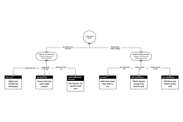
Plant Cell or Animal Cell — What Tells Them Apart?
Plant and animal cells share almost everything. Only three structures are found in a plant cell and never in an animal one, and the shared parts settle nothing. Sort six structures in an interactive playground.
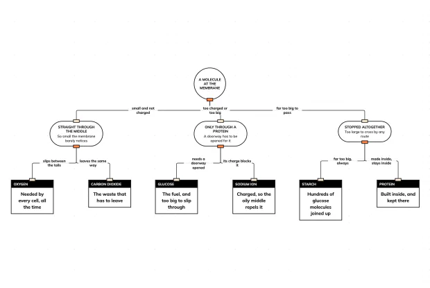
What Does the Cell Membrane Let Through?
A cell membrane is a double layer of fat molecules with proteins set into it. Small uncharged molecules cross it freely, charged or larger ones need a protein doorway, and big ones never get through. Sort six molecules in an interactive playground.
What Does the Nucleus Actually Hold?
The nucleus holds DNA, but chromosome, chromatin and gene all describe that same material at different levels. Follow the zoom from a cell down to a single gene in an interactive playground.
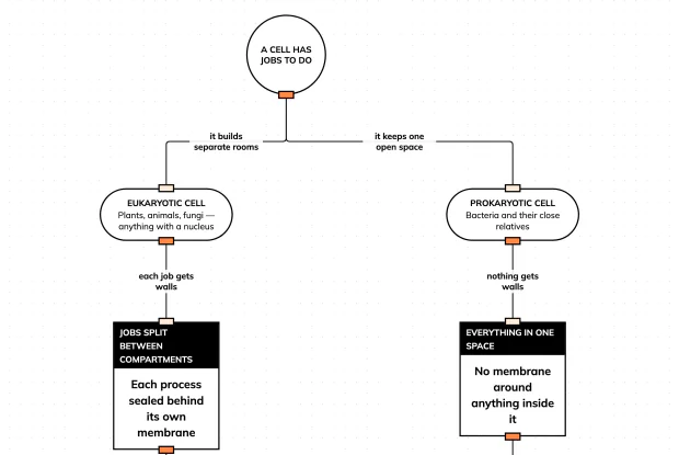
Bacteria Have No Nucleus — So What Do They Have?
A bacterium has no nucleus and no membrane-bound organelles, yet it does everything a cell must do. Follow the two ways of organising a cell in an interactive playground.