How Does One Cell Become Two? Mitosis in Order
Root Concept
In mitosis a cell copies every chromosome, the copies condense and line up across the middle of the cell attached to fibres from both ends, the pairs are pulled apart so that a complete set reaches each end, and the cell then divides into two genetically identical cells.
CodePLU Goal
Upgrading Human Mental Models
Learn how to think in Workflows

Concept Development By codeplu.com
Every stage is about one thing: making sure each side ends up with a complete set.
The Hardest Part Is Not the Splitting
A cell dividing into two sounds like a problem of cutting something in half, and for the cytoplasm it more or less is — the cell pinches in and the contents are shared out, and precision hardly matters. A cell with slightly more or slightly less cytoplasm than its sister will manage perfectly well.
The chromosomes are a different matter entirely. Each new cell needs a complete set, not roughly half the material. A cell that ends up with two copies of one chromosome and none of another is in serious trouble, and no amount of extra cytoplasm will make up for it. So the whole process is organised around solving that problem, and the stages in the playground are the solution.
The method has two clever parts. First, the DNA is copied before anything is separated, so that each chromosome exists as two identical copies held together. Second — and this is the step people skip — the copies are brought to the centre of the cell and lined up in a single plane, with fibres attached to each pair reaching out to opposite ends. Only once everything is lined up and attached are the pairs pulled apart. That ordering is what makes the result reliable: because every pair is attached to both ends before anything moves, one copy of every chromosome is guaranteed to go each way.
This is how growth happens, how damaged tissue is repaired, and how every asexually reproducing organism in the previous tutorial makes more of itself.
Why the Stages Are in This Order
Why copy the DNA before dividing anything?
Because you cannot share out one set of chromosomes between two cells and give each a complete set. There is only one of each to begin with, so the material has to be doubled first and the two cells then take one copy each. The copying happens well before the visible stages of division begin, during the long stretch of a cell's life when it is simply going about its business. After it, each chromosome exists as two identical copies joined together at a point along their length, which is why a chromosome drawn in a textbook so often has the familiar X shape. That shape is not what a chromosome normally looks like — it is what one looks like after it has been copied and before it has been pulled apart.
Why do the chromosomes only become visible now?
Because for most of a cell's life the DNA is unwound into extremely long, fine threads, far too thin to see and far too tangled to move around. In that state the genes can be read, which is the normal business of a cell. Before division the threads coil up on themselves repeatedly until each chromosome is a short thick structure, which does two things: it makes them compact enough to be hauled about without tangling or breaking, and it incidentally makes them thick enough to see under a light microscope. This is why the tutorial on the nucleus noted that chromosomes appear only when a cell divides — they are present the whole time, and merely in a form nothing can see.
What is the lining-up step actually for?
It is the step that makes the whole thing accurate, and it is the one most often passed over. The copied chromosomes are brought to the middle of the cell and arranged in a single plane, and fibres extend from both ends of the cell and attach to each pair — one fibre to each copy, pulling in opposite directions. Nothing separates until every pair is attached on both sides. Because of that, when the pulling starts, one copy of every chromosome necessarily goes to each end. The accuracy is not a matter of care during the separation; it is built into the arrangement made beforehand, which is a far more reliable way of getting something right than checking afterwards.
Where does mitosis happen in an organism?
Wherever new cells are needed, which is a great deal of the time. It is how a fertilised egg becomes an embryo and then an organism, so every cell in your body traces back through mitosis to a single cell. It is how you grow. It is how a cut heals and how the lining of your gut and the surface of your skin are continually replaced. In plants it is concentrated in the meristems at the tips and in the cambium, which an earlier track located, since those are the only places a plant makes new cells at all. And it is how every organism on the asexual branch of the previous tutorial reproduces.
Real World Example
Looking at a Root Tip Under a Microscope
A root tip is the standard specimen for seeing mitosis, and the reason it works is a direct consequence of where plants grow from.
Step 1
The tip is chosen because of the meristem
An earlier track established that a plant grows only at its tips and from a ring inside the stem, and that everywhere else is permanent tissue whose cells have stopped dividing. That makes the few millimetres at the end of a root the single best place to look for cells caught in the act of division, because it is one of the only places where division is happening at all. A root tip from a germinating onion or broad bean, which can be grown on a windowsill in a jar of water, is the usual specimen.
Step 2
The tissue is softened and stained
The tip is cut off and warmed in dilute hydrochloric acid, which breaks down the substance gluing the cells together so that they can be spread into a single layer. Without that step the cells sit in several layers on top of one another and nothing can be seen clearly. A stain is then added that binds to DNA and makes the chromosomes stand out — several are used, and most turn them a strong red or purple. The tip is squashed flat under a cover slip and the slide is ready, with the cells spread thinly enough for light to pass through a single layer of them.
Step 3
Every stage appears somewhere in the field
What you see is not one cell going through the sequence but hundreds of cells frozen at different points in it, because the specimen was killed at a single instant and every cell was somewhere different. Most show no chromosomes at all, since cells spend most of their time between divisions. A minority show the coiled chromosomes, some lined up neatly across the middle, some pulled into two groups at opposite ends. Counting how many cells are at each stage gives a rough measure of how long each stage lasts, since a stage that takes longer catches more cells in it.
FAQs
Final Words
Six stages, and every one of them exists to solve the same problem: making sure each new cell gets a complete set of chromosomes rather than roughly half the material. The DNA is copied first, because one set cannot be shared. The copies coil up, because long fine threads cannot be moved without tangling. They line up in the middle attached to fibres from both ends, and only then are they pulled apart.
That ordering is the accuracy. Nothing separates until everything is attached on both sides, so one copy of every chromosome is guaranteed to go each way, and the two cells that result are genetically identical. This is how growth and repair happen and how every organism on the asexual branch reproduces. The next tutorial asks what has to change when two cells are going to fuse.
Continue This Track
This concept is part 2 of How Living Things Make More of Themselves.
One Parent or Two? The Two Ways to Reproduce
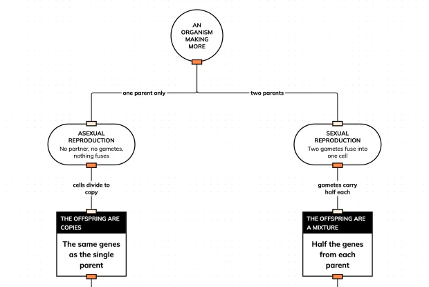
One parent gives copies, quickly. Two parents give offspring that all differ, slowly. Follow both routes from the same starting point in an interactive playground and see what each one buys.
How Does One Cell Become Two? Mitosis in Order
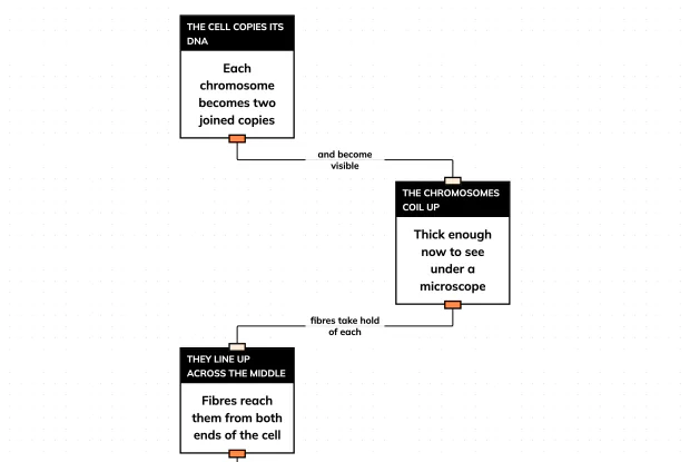
Mitosis makes two cells with identical sets of chromosomes. Follow the six stages in order in an interactive playground, and see why the lining-up step is what makes it accurate.
Why Do Gametes Carry Half? Follow the Arithmetic
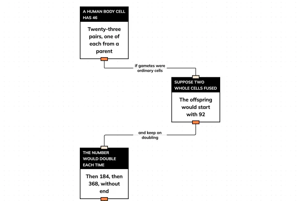
If a sperm and an egg each carried a full set of chromosomes, every generation would have twice as many as the last. Reason to meiosis step by step in an interactive playground.
Making More Without a Partner — Four Methods
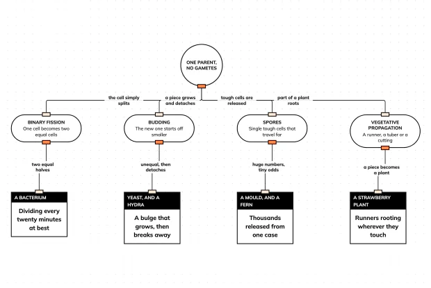
Asexual reproduction is not one process. Sort binary fission, budding, spores and vegetative propagation by how the new organism actually gets started, in an interactive playground.
Why Have Two Parents At All? Variation Is Why
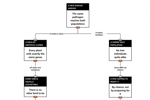
Sexual reproduction is slow, expensive and needs a partner. Trace what happens when one disease meets a clone population and a varied one, in an interactive playground, and see what the cost buys.